
Intradermal Nevus: When a Raised Mole Is Harmless and When to Monitor

Medically reviewed by Oghenefejiro Okifo | MD, Harvard Medical School | Henry Ford Hospital - Detroit, MI on April 16th, 2026.
Published on April 9th, 2026. Updated on July 23rd, 2026.
Key takeaways
An intradermal nevus is a mature, benign mole where melanocytes have migrated deep into the dermis, giving it a soft, dome-shaped, flesh-colored or light brown appearance.
Intradermal nevi are among the most common skin growths in adults and carry very low malignant potential compared to other types of moles.
The ABCDE rule (Asymmetry, Border, Color, Diameter, Evolution) applies to any mole, including intradermal nevi, to catch early warning signs.
Cosmetic removal through shave excision, punch biopsy, or laser treatment is a quick outpatient procedure with minimal downtime.
Rapid growth, color change, irregular borders, bleeding, or itching in any raised mole warrant prompt dermatologist evaluation.
What Makes a Mole "Intradermal"
An intradermal nevus is a benign, raised mole where melanocytes have fully migrated into the deeper dermis, leaving it soft, dome-shaped, and often flesh-colored or pale. Most adults have between 10 and 40 moles, and not all are the same type.
Because the melanocytes are no longer at the surface, the mole loses much of its pigment over time, becoming a raised, soft bump that is flesh-colored, light pink, or pale tan. These moles feel smooth or slightly warty and range from a few millimeters to about a centimeter. Recognizing what is normal for this type of growth makes it easier to notice when something changes.
How an Intradermal Nevus Differs from Other Moles
Moles follow predictable developmental stages.
Junctional Nevus: The Starting Point
A junctional nevus forms when melanocytes cluster at the junction between the epidermis and dermis. These moles are flat, evenly pigmented, and dark brown or black, most common in children and young adults.
Compound Nevus: The Middle Stage
A compound nevus has melanocytes at the junction and within the dermis, resulting in a slightly raised center and a flat outer edge. These brown-toned moles are common in teenagers and adults through their thirties.
Intradermal Nevus: The Mature Stage
As a compound nevus ages, the junctional component fades, and melanocytes move fully into the dermis. The mole loses pigmentation and rises off the surface. Many people notice these pale, raised bumps appearing on the face, neck, and scalp during middle age and beyond.
A raised, pale bump that has remained unchanged for years is almost always an intradermal nevus.
Intradermal Melanocytic Nevus: What Makes It Different
The full clinical name for this type of mole is intradermal melanocytic nevus, and dermatologists use this term specifically to describe a benign lesion made up of mature melanocytes that have completed their downward migration into the dermis. Understanding what that means in practice can help you feel confident the next time a doctor uses the term in your chart notes.
Melanocytes are the cells responsible for producing melanin, the pigment that gives skin and moles their color. In earlier mole types, such as junctional and compound nevi, these cells remain active near or at the skin's surface. In an intradermal melanocytic nevus, the cells have settled deep in the dermis and are no longer dividing actively. This is why the mole loses its dark color over time and becomes flesh-toned or pale pink.
How It Looks Under the Microscope
On a pathology report, an intradermal melanocytic nevus shows nests of melanocytes entirely within the dermis, with no involvement of the dermal-epidermal junction. The cells are typically small, evenly spaced, and show no signs of atypical nuclear features. Pathologists describe this pattern as "maturation," meaning the cells have aged in a predictable, controlled way. This microscopic appearance is what gives dermatologists confidence that the growth is benign.
Who Gets Intradermal Melanocytic Nevi
These lesions are most common in adults between the ages of 30 and 60. They tend to appear on the face, neck, scalp, and upper trunk. Sun-exposed skin is the most common site, though they can develop anywhere on the body. People with lighter skin tones tend to notice them more easily because the pale bump contrasts with surrounding skin, but they occur across all skin types.
Distinguishing It from Other Growths
An intradermal melanocytic nevus is frequently mistaken for other common benign growths, including dermatofibromas, basal cell carcinomas, and skin tags. The key distinguishing features are its soft, compressible texture, its pale or flesh-toned color, and its gradual appearance over many years without rapid change. A dermatologist can usually distinguish these growths with a dermatoscope during a routine office visit. If there is any doubt, a simple biopsy settles the question definitively and provides a tissue sample for pathology review.
Why Intradermal Nevi Are Almost Always Benign
The deep dermal location of melanocytes is the key reason intradermal nevi carry very low malignant potential. Melanoma most commonly arises from junctional activity, where surface melanocytes are actively dividing and exposed to UV damage. In an intradermal nevus, those cells have already migrated away from the surface and are no longer in an active growth phase.
Malignant transformation is rare enough that most dermatologists manage these moles through routine observation rather than prophylactic removal. That said, low risk is not no risk, which is why monitoring still matters.
The ABCDE Rule: Monitoring Any Mole
The ABCDE rule is the standard framework dermatologists recommend for evaluating all moles , including raised intradermal nevi.
Asymmetry: Draw an imaginary line through the center. In a benign mole, both halves look roughly the same. An asymmetric mole, where one side looks very different from the other, warrants attention.
Border: Benign moles have smooth, clearly defined edges. Ragged, notched, or blurred borders that fade into surrounding skin are a concern.
Color: A healthy mole is one uniform color, whether dark brown, light tan, or even flesh-colored. Multiple colors within a single mole, including shades of red, white, blue, or black, call for evaluation.
Diameter: Most benign moles are smaller than 6 millimeters, roughly the diameter of a pencil eraser. Moles larger than this should be documented and monitored.
Evolution: This is arguably the most critical criterion. Any mole that changes in size, shape, color, or texture over weeks or months needs professional evaluation. New symptoms like itching, bleeding, or crusting also count as evolution.
For intradermal nevi specifically, the E criterion matters most. These moles should look the same year after year. A raised bump on your face that has been stable for a decade is reassuring. One that has recently grown, darkened, started catching on clothing, or is bleeding needs to be seen by a dermatologist.
When to Worry About a Raised Mole or Intradermal Melanocytic Nevus
Most intradermal nevi are genuinely benign and require only routine self-monitoring. Certain changes should prompt a visit to a dermatologist without delay.
Rapid or sudden growth is a red flag regardless of a mole's history. A bump that has been stable for years and then doubles in size over a few months is not behaving normally.
Color change in any direction deserves attention. An intradermal nevus becoming darker, developing new pigmented areas, or showing multiple colors is unusual and warrants evaluation.
Irregular borders that were not there before suggest new cellular activity. If a mole's edge becomes notched, blurry, or uneven, that is worth examining.
Bleeding from a mole that has not been injured is always a reason to see a doctor. Occasionally, scratching or bumping a raised mole causes minor bleeding that resolves quickly, but spontaneous or recurrent bleeding requires evaluation.
Persistent itching or tenderness without an obvious cause, like friction or an insect bite, should be assessed. These sensations can accompany inflammatory or proliferative changes within a mole.
See a doctor online today
A licensed doctor can review your symptoms online, talk through next steps, and send a prescription to your pharmacy if appropriate.
- $39 video visit
- Insurance accepted
- 4.8 on Trustpilot
How Dermatologists Evaluate Moles: Dermatoscopy
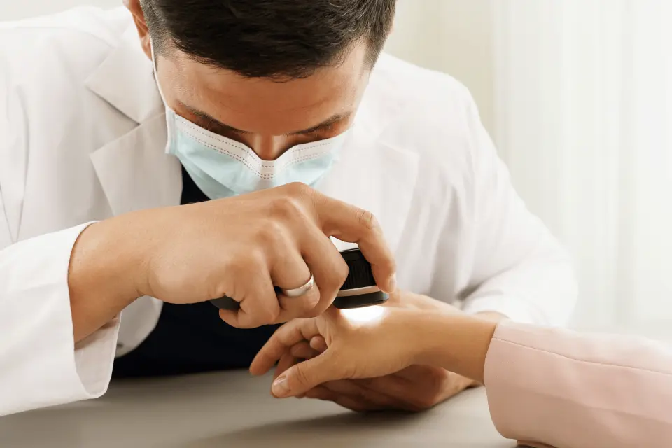
When you visit a dermatologist with a concerning mole, the evaluation will almost always involve dermatoscopy, a handheld magnifying device with polarized light that reveals architectural patterns invisible to the naked eye.
Benign intradermal nevi show characteristic dermatoscopic patterns: comma-shaped vessels, globular structures, and no irregular pigment networks. If suspicious features appear, the dermatologist will recommend a biopsy: shave (removing the raised portion with a blade), punch (a cylindrical core sample), or excisional (removing the mole with a margin). A pathologist then confirms the diagnosis.
Raised moles can also look similar to seborrheic keratoses, another common benign growth. Knowing how to tell a seborrheic keratosis from melanoma helps reduce unnecessary worry and ensures the right growths get professional attention.
Cosmetic Removal Options
Intradermal nevi are benign, but many people choose to remove them for cosmetic reasons or because the location causes practical problems, such as a mole on the neck that gets irritated by collars or a facial bump that catches on glasses.
Shave excision is the most common method. The dermatologist numbs the area with a small injection of local anesthetic and uses a surgical blade to shave the raised portion of the mole flush with the skin. The procedure takes about ten minutes in the office, requires no stitches, and leaves a small pink mark that fades over several weeks.
Punch biopsy is used for smaller intradermal nevi. A circular blade removes the mole and a small margin of surrounding tissue. This technique may leave a tiny scar but provides a complete tissue sample for pathology if there is any diagnostic uncertainty.
Laser treatment, particularly with ablative lasers, can vaporize the tissue of an intradermal nevus. This approach works well for flat or only slightly raised moles on cosmetically sensitive areas. It does not provide a tissue sample, so it is appropriate only when the mole has been clinically confirmed as benign before treatment.
All three methods are outpatient procedures performed under local anesthesia. Recovery involves keeping the area clean and protected from sun exposure for a few weeks. Recurrence is possible with any method, particularly if cells remain at the base of the lesion after shave excision.
If you are considering removal, telehealth can be a convenient first step to determine whether a mole is appropriate for a cosmetic procedure or requires a diagnostic biopsy first. Doctronic.ai can help you frame the right questions before your in-person appointment.
Performing Skin Self-Exams
Monthly self-examination is the most practical tool for detecting early changes in moles. Stand in a well-lit room with a full-length mirror. Examine the front and back of your body, raise your arms to check your sides, and use a hand mirror to inspect your scalp and the backs of your ears. Check your feet, including between your toes and the soles.
Photograph any moles that concern you to create a baseline record. Comparing a photo from six months ago to today is far more reliable than relying on memory.
Annual full-body skin examinations by a dermatologist add professional assessment to your personal monitoring. People with many moles, a personal or family history of skin cancer, or fair skin that burns easily may benefit from more frequent professional exams.
Frequently Asked Questions
An intradermal nevus is almost always benign. The melanocytes in this type of mole have matured and moved deep into the dermis, where they are no longer in an active growth phase. Malignant transformation is rare, but any mole that changes in size, shape, color, or texture should be evaluated by a dermatologist.
An intradermal nevus typically appears as a soft, dome-shaped bump that is flesh-colored, light pink, or pale tan. It may feel smooth or slightly warty and usually ranges from a few millimeters to about a centimeter in size. These moles often develop on the face, neck, and scalp in middle age and beyond.
Removal is not medically necessary for a confirmed intradermal nevus, but many people choose to remove them for cosmetic reasons or because the location causes irritation. Options include shave excision, punch biopsy, and laser treatment, all performed as quick outpatient procedures. Our AI doctor can help you figure out whether your mole is a good candidate for cosmetic removal or needs a diagnostic biopsy first.
An intradermal nevus is a benign mole with mature, non-dividing melanocytes deep in the dermis, while melanoma is a malignant skin cancer that arises from active, dividing melanocytes. Key warning signs that a raised mole may not be benign include rapid growth, multiple colors, irregular borders, and spontaneous bleeding. A dermatologist can evaluate any concerning mole with dermatoscopy and, if needed, a biopsy.
For stable, unchanged moles, a yearly full-body skin exam with a dermatologist is generally sufficient. Monthly self-exams using the ABCDE rule help you spot any changes between professional visits. People with many moles, a personal or family history of skin cancer, or fair skin may benefit from more frequent professional evaluations.
The Bottom Line
An intradermal nevus is a mature, benign mole that forms when melanocytes migrate deep into the dermis, leaving a soft, raised, flesh-colored bump that is almost always harmless. Understanding this type of mole, how it differs from junctional and compound nevi, and what changes to watch for puts you in a much stronger position to protect your skin health. The ABCDE rule and monthly self-exams remain the most reliable tools for catching anything unusual early. When a raised mole grows, changes color, bleeds, or develops irregular borders, a dermatologist visit is the right next step. For quick guidance on whether a skin change needs professional evaluation, visit Doctronic.ai for AI-powered consultations available any time of day.
References
AAD: 'Moles' Northwell Health: '4 Reasons You Need to See a Dermatologist'
Related articles
What Is a Dangerously High Platelet Count Level and What Does It Mean?
What Is a Dangerously Low Platelet Count Level and What Does It Mean?
What Is a Dangerously Low White Blood Cell Count Level and What Does It Mean?
New Sickle Cell Treatments 2026: Latest FDA Approvals and Breakthrough Therapies
Get personalized health advice
Chat Now