
Stage 1 Melanoma: Diagnosis, Treatment, and Survival Rates
What Stage 1 Melanoma Means for PatientsA stage 1 melanoma diagnosis is understandably alarming, but the prognosis at this early stage is overwhelmingly positive. The cancer [...]
Read More
Medically reviewed by Alan Lucks | MD , Alan Lucks MDPC Private Practice - New York on May 24th, 2026. Updated on May 28th, 2026
Stage 1 melanoma has an excellent prognosis, with five-year survival rates around 99 percent when caught early
The distinction between Stage 1A and Stage 1B depends on tumor thickness and whether ulceration is present
Wide local excision surgery remains the primary treatment, with specific margin requirements based on tumor characteristics
Sentinel lymph node biopsy may be recommended for Stage 1B cases to check for microscopic spread
Long-term monitoring and sun protection are essential for preventing recurrence and detecting new melanomas
Doctronic.ai offers telehealth visits to help evaluate suspicious skin changes and guide next steps
A stage 1 melanoma diagnosis is understandably alarming, but the prognosis at this early stage is overwhelmingly positive. The cancer remains localized to the skin and has not spread to lymph nodes or distant organs. When caught at this stage, five-year survival exceeds 99 percent, making early detection the single most important factor in outcomes. Understanding what stage 1 means, how it is treated, and what monitoring looks like afterward helps patients navigate the experience with clarity.
The difference between Stage 1A and Stage 1B comes down to two measurable factors: tumor thickness and ulceration status. Stage 1A melanomas are less than 0.8 millimeters thick with no ulceration, meaning the tumor surface remains intact. Stage 1B melanomas are either less than 0.8 millimeters thick with ulceration, or between 0.8 and 1.0 millimeters thick with or without ulceration.
This distinction matters because it directly influences treatment planning, surgical margin requirements, and the recommendation for sentinel lymph node biopsy. Stage 1B carries a slightly higher risk profile, though both substages have excellent long-term outcomes.
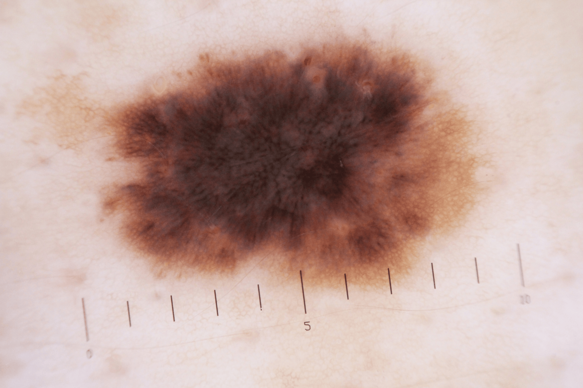
Breslow depth, measured in millimeters by a pathologist, indicates how deeply the melanoma has grown into the skin. Thinner tumors generally carry a better prognosis. Ulceration, in which the tumor has broken through the overlying skin, indicates more aggressive behavior regardless of thickness. Ulcerated melanomas of the same depth as non-ulcerated ones tend to have slightly higher recurrence rates, which is why pathology reports always note both factors.
When a dermatologist identifies a suspicious lesion, an excisional biopsy is the preferred initial diagnostic step. This involves removing the entire visible lesion along with a small margin of surrounding skin. Punch or shave biopsies may be used in some situations, but can underestimate tumor thickness if the deepest portion is not captured.
The pathology report from a biopsy provides critical information, including Breslow depth, ulceration status, mitotic rate, and margin status. These details determine the melanoma's precise stage and guide all subsequent treatment decisions.
The ABCDE rule provides a practical framework for identifying potentially dangerous moles: Asymmetry, Border irregularity, Color variation, Diameter larger than a pencil eraser, and Evolving appearance. Regular self-examinations using these criteria, combined with annual professional skin checks, catch melanomas at their most treatable stage.
Sentinel lymph node biopsy may be recommended for Stage 1B melanomas, particularly those 1.0 millimeters or thicker, or between 0.8 and 1.0 millimeters with ulceration. The procedure uses a radioactive tracer and blue dye to identify the first lymph node where cancer cells would travel if they spread. A negative result confirms the melanoma remains localized, while a positive result changes the staging and treatment approach.
Surgery is the standard treatment for stage 1 melanoma, and most patients need no additional treatment beyond the procedure. Wide local excision removes the melanoma site along with a margin of healthy tissue to ensure no cancer cells remain. The procedure is typically performed as an outpatient surgery under local anesthesia using an elliptical removal pattern. Tissue is sent to pathology to confirm clear margins.
Margin requirements depend on tumor thickness. Melanomas less than 1 millimeter thick generally require 1-centimeter margins, while those between 1 and 2 millimeters thick may need 1 to 2-centimeter margins. The surgical site may be tender for one to two weeks, and stitches are typically removed within two weeks. Most patients return to normal activities within days, though strenuous exercise may need to wait until healing is complete.
Stage 1 melanoma carries five-year survival rates exceeding 98 to 99 percent, with Stage 1A having a slightly better outlook than Stage 1B. Both substages maintain survival rates above 90 percent at ten years. These numbers reflect the effectiveness of early surgical intervention and underscore why screening programs emphasize catching melanoma before it advances. Several factors influence individual prognosis beyond staging, including age, tumor location (trunk and head-neck sites carry a marginally higher risk), immune health, and family history of melanoma.
After treatment, dermatologists typically recommend full-body skin examinations every three to six months for the first two years, then every six to twelve months thereafter. Monthly self-examinations between appointments help catch any changes early. Recognizing what early melanoma looks like and monitoring existing moles for changes remains important long after initial treatment.
Sun protection becomes a permanent priority after a melanoma diagnosis. Applying broad-spectrum SPF 30 or higher daily, seeking shade during peak UV hours, wearing protective clothing, and completely avoiding tanning beds reduces the risk of both recurrence and new melanoma development. Monitoring vitamin D levels through regular blood work helps ensure that sun avoidance does not lead to nutritional deficiencies.
Most patients recover within two to three weeks. The surgical site may feel tender initially, but the procedure is minimally invasive and performed under local anesthesia. Strenuous activities should be avoided until the incision heals completely.
Stage 1 melanoma has a recurrence rate of approximately five percent or fewer. Regular monitoring detects any recurrence early, helping maintain the excellent prognosis associated with early-stage disease.
Chemotherapy is not part of standard treatment for stage 1 melanoma. Surgery alone is typically sufficient, and additional systemic treatments are reserved for more advanced stages.
A positive result indicates that melanoma cells have reached the nearest lymph node, which changes the staging to 3A or higher and may warrant additional treatment options, including immunotherapy or closer surveillance.
Stage 1 melanoma carries an excellent prognosis when detected and treated promptly, with five-year survival rates exceeding 99 percent. Surgery is the primary treatment, and long-term monitoring with consistent sun protection minimizes the risk of recurrence. For concerns about suspicious moles or skin changes, Doctronic.ai offers telehealth visits to help evaluate symptoms and determine whether in-person dermatology care is needed.
What Stage 1 Melanoma Means for PatientsA stage 1 melanoma diagnosis is understandably alarming, but the prognosis at this early stage is overwhelmingly positive. The cancer [...]
Read More
Join 50,000+ readers using Doctronic to understand symptoms, medications,
and next steps.
Add your phone number below to get health updates and exclusive VIP offers.
By providing your phone number, you agree to receive SMS updates from Company. Message and data rates may apply. Reply “STOP” to opt-out anytime. Read our Privacy Policy and Terms of Service for more details.
Save your consults. Talk with licensed doctors and manage your health history.