
Differin (Adapalene) for Seniors: What to Know
Read More

Medically reviewed by Veronica Hackethal | MD, MSc , Harvard University | University of Oxford | Columbia Vagelos College of Physicians and Surgeons on May 25th, 2026. Updated on May 26th, 2026
Annual skin checks by a dermatologist catch cancers that self-exams often miss, with about 8,500 people diagnosed with skin cancer daily in the U.S.
The ABCDE method helps identify melanoma warning signs: Asymmetry, Border irregularity, Color variation, Diameter over 6mm, and Evolution of moles
Non-melanoma skin cancers like basal cell and squamous cell carcinoma have distinct appearances that trained dermatologists recognize quickly
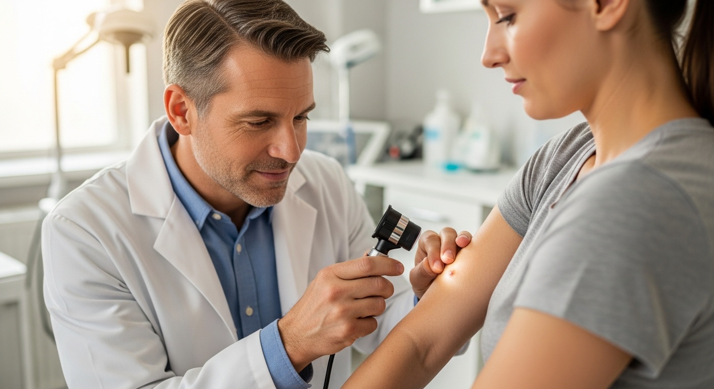
Dermoscopy and total body photography provide detailed views of suspicious spots that naked-eye exams cannot detect
If a concerning spot appears, a simple biopsy determines whether treatment is needed, often catching problems before they spread
Have questions about a suspicious mole or skin change? Doctronic.ai offers free AI doctor visits and affordable telehealth consultations to evaluate your concerns quickly
Your bathroom mirror cannot save your life. Self-exams have value, but they miss what trained dermatologists catch routinely during professional skin screenings. The statistics are stark: 1 in 5 Americans will develop skin cancer in their lifetime, making this the most common cancer in the country. When a dermatologist examines your skin during an annual check, they bring years of pattern recognition, specialized tools, and medical knowledge that no app or mirror can replicate.
Understanding what happens during these exams removes the mystery and helps patients prepare. A dermatologist looks for specific warning signs, uses particular techniques, and follows established protocols when something appears suspicious. Doctronic.ai offers resources to help patients understand skin health between appointments, but nothing replaces hands-on professional evaluation. Knowing what your dermatologist searches for empowers you to become a better partner in protecting your own skin.
Self-checks matter, but they have serious limitations. Most people cannot see their own backs, scalps, or the soles of their feet properly. Even visible areas present challenges because untrained eyes often cannot distinguish between harmless spots and dangerous lesions. Dermatologists spend years learning subtle differences that determine whether a mark requires immediate attention or simple monitoring.
Professional skin cancer screening detects cancer early, monitors existing moles for changes, and identifies precancerous lesions before they become dangerous. Early-stage melanoma has a five-year survival rate of about 99% when detected before metastasis. That number drops dramatically once cancer reaches lymph nodes or other organs.
Not everyone faces equal skin cancer risk. Fair skin, light eyes, freckling tendency, and history of sunburns increase vulnerability significantly. Family history of melanoma, previous skin cancer diagnosis, or having many moles also raises concern. People who used tanning beds, especially before age 35, face substantially higher melanoma risk.
Your dermatologist considers these factors when deciding examination frequency and intensity. Someone with multiple risk factors may need checks every six months rather than annually. Doctronic.ai can help patients assess their personal risk profiles and understand when professional evaluation becomes urgent.
Dermatologists use the ABCDE system as a primary screening tool. Asymmetry means one half of a mole does not match the other half. Normal moles tend toward symmetry, while melanomas often grow unevenly. Border irregularity refers to edges that appear ragged, notched, or blurred rather than smooth and well-defined.
These characteristics signal abnormal cell growth patterns. When cells multiply in disorganized ways, the resulting lesion reflects that chaos visually. Your dermatologist examines each mole systematically, comparing halves and tracing borders with trained precision.
Healthy moles typically display uniform coloring throughout. Melanomas frequently show multiple shades of brown, tan, black, red, white, or blue within a single lesion. This color variation indicates different cell populations growing at different rates, a hallmark of malignancy.
Diameter matters too. Moles larger than 6 millimeters, roughly the size of a pencil eraser, warrant closer examination. Size alone does not confirm cancer, but larger lesions provide more opportunity for dangerous changes to develop unnoticed.
The "E" in ABCDE may be most important. Any mole that changes size, shape, color, elevation, or develops new symptoms like bleeding, itching, or crusting requires immediate professional evaluation. Stable moles rarely cause problems. Changing moles demand attention.
This is why annual checks matter: dermatologists compare current appearances to previous examinations. They notice subtle shifts that patients themselves miss because changes happen gradually over months.
Basal cell carcinoma is the most common skin cancer, appearing as pearly or waxy bumps, flat flesh-colored or brown lesions, or bleeding sores that heal and return. These cancers grow slowly and rarely spread to distant organs, but they can cause significant local damage if ignored.
Dermatologists recognize the characteristic "rolled edges" and visible blood vessels that distinguish basal cell carcinoma from harmless bumps. Early removal prevents disfiguring tissue destruction.
Squamous cell carcinoma presents as firm red nodules, flat lesions with scaly crusted surfaces, or sores that heal and reopen repeatedly. Unlike basal cell carcinoma, squamous cell can spread to lymph nodes and other organs if left untreated.
Sun-exposed areas face highest risk: face, ears, neck, lips, and backs of hands. Your dermatologist pays particular attention to these zones during examinations.
Actinic keratoses are rough, scaly patches caused by years of sun exposure. These are not yet cancer but can progress to squamous cell carcinoma over time. Dermatologists often treat these preventively through freezing, topical medications, or other methods.
Finding and treating actinic keratoses stops skin cancer before it starts. This preventive approach represents one of the most valuable aspects of annual skin checks.
Dermoscopy uses a handheld device with magnification and specialized lighting to examine skin structures invisible to the naked eye. The dermatologist sees pigment patterns, blood vessel arrangements, and cellular structures that reveal whether a lesion appears benign or suspicious.
This technique dramatically improves diagnostic accuracy. Studies show dermoscopy helps dermatologists detect melanomas earlier and avoid unnecessary biopsies of harmless moles.
For high-risk patients, total body photography creates a baseline record of all moles and marks. Subsequent visits compare new images to previous ones, making even subtle changes obvious. Digital mole mapping tracks specific lesions over time with precise measurements.
These technologies transform skin cancer screening from snapshot evaluation to longitudinal monitoring. Changes that might escape notice during a single exam become apparent when compared across multiple sessions.
When a dermatologist identifies a concerning lesion, biopsy provides definitive answers. The procedure is simple: after numbing the area with local anesthetic, the doctor removes all or part of the suspicious tissue. Most biopsies take minutes and heal within two weeks.
Different biopsy types serve different purposes. Shave biopsies remove surface layers, punch biopsies extract deeper circular samples, and excisional biopsies remove entire lesions. Your dermatologist selects the appropriate technique based on the lesion's characteristics.
A pathologist examines biopsy tissue under a microscope and provides a detailed report. Results typically arrive within one to two weeks. The report specifies whether cancer exists, what type, how deep it extends, and whether margins are clear.
Doctronic.ai helps patients understand pathology terminology and prepare questions for follow-up appointments. Clear communication between patients and dermatologists ensures appropriate treatment decisions.
Daily sun protection remains the single most effective prevention strategy. Use broad-spectrum SPF 30 or higher sunscreen, wear protective clothing, seek shade during peak UV hours, and avoid tanning beds entirely. Monthly self-exams complement annual professional checks by catching obvious changes between appointments.
Document new or changing spots with photographs. Note any moles that itch, bleed, or develop crusting. Bring these observations to your next dermatology appointment for professional evaluation.
Most comprehensive skin examinations take 10 to 15 minutes. Patients with many moles or previous skin cancer history may require longer appointments for thorough evaluation.
Yes. Melanoma can develop under fingernails and toenails. Clear nails allow your dermatologist to examine these often-overlooked areas completely.
Wear loose, easy-to-remove clothing. You will change into a gown for a full-body examination. Remove jewelry and accessories that might obscure skin areas.
People with previous skin cancer, many atypical moles, or strong family history may need examinations every three to six months rather than annually.
Absolutely. Screening examinations catch problems before symptoms appear. Many skin cancers cause no pain or discomfort in early stages.
Annual skin checks save lives by catching skin cancer when treatment works best. With about 8,500 daily skin cancer diagnoses in the U.S., professional screening is essential protection. Visit Doctronic.ai for AI-powered health guidance and convenient telehealth appointments to discuss skin concerns with licensed doctors anytime.

Join 50,000+ readers using Doctronic to understand symptoms, medications,
and next steps.
Add your phone number below to get health updates and exclusive VIP offers.
By providing your phone number, you agree to receive SMS updates from Company. Message and data rates may apply. Reply “STOP” to opt-out anytime. Read our Privacy Policy and Terms of Service for more details.
Save your consults. Talk with licensed doctors and manage your health history.