Early stage melanoma has a five-year survival rate around 99% when caught before it spreads beyond the skin.
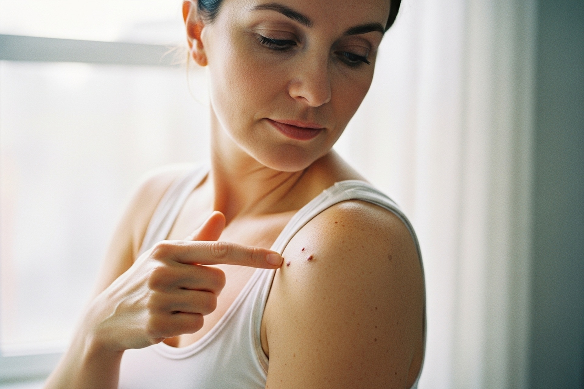
The ABCDE method helps identify suspicious moles: Asymmetry, Border irregularity, Color variation, Diameter over 6mm, and Evolving characteristics.
Melanoma can hide in unexpected places like nail beds, palms, and soles of feet.
Monthly self-exams combined with annual professional screenings offer the best protection against late diagnosis.
About 70% of melanomas arise as new spots rather than from existing moles, making whole-body monitoring essential.
Doctronic.ai provides AI-powered consultations to help evaluate skin concerns and determine when a dermatology appointment is needed.
Why Catching Early Stage Melanoma Saves Lives
A small dark spot on the skin might seem harmless. Most of the time, it is. But when that spot turns out to be melanoma, timing becomes everything. Melanoma is the deadliest form of skin cancer, yet it is also one of the most treatable when found early.
Approximately 103,000 new cases of melanoma will be diagnosed in the U.S. in 2026, making awareness more critical than ever. The melanoma survival rates drop dramatically once the cancer advances past its earliest stages.
Understanding Stage 0 and Stage I Melanoma
Stage 0 melanoma, called melanoma in situ, means cancer cells exist only in the top layer of skin. They have not invaded deeper tissue. Stage I melanoma has grown slightly deeper but remains localized without spreading to lymph nodes or distant organs.
Both stages share one important characteristic: the cancer is still contained. A dermatologist can often remove Stage 0 melanoma completely with a simple excision. Stage I typically requires a wider surgical margin but still carries an excellent prognosis.
The Gap Between Early and Late Detection
Stage 0 and Stage I melanoma have five-year survival rates ranging between 97% and 99%. Once melanoma reaches Stage IV, that number drops to around 32%.
This gap exists because early melanoma has not yet entered the bloodstream or lymphatic system. Surgical removal at this point eliminates the threat entirely for most patients. Doctronic.ai helps people understand their symptoms and determine when professional evaluation is needed.
Visual Warning Signs: The ABCDE Method
The ABCDEs of melanoma provide a practical framework for evaluating suspicious moles at home.
Asymmetry and Irregular Borders
Healthy moles are typically round or oval with even halves. Melanoma often grows unevenly, creating asymmetry where one side looks different from the other.
Border irregularity provides another red flag. Normal moles have smooth, well-defined edges. Melanoma borders tend to be ragged, notched, or blurred, fading into surrounding skin without a clear stopping point.
Color Variations and Diameter
A single mole should not contain multiple colors. Melanoma often displays shades of brown, black, tan, red, white, or blue within the same lesion. This color variation happens because cancer cells grow at different rates and depths.
Size matters too. Most melanomas are larger than 6 millimeters in diameter, roughly the size of a pencil eraser. Smaller melanomas exist, but any mole that continues growing deserves evaluation.
Evolving Moles: The Most Important Warning Sign
Many dermatologists consider evolution the most critical indicator. Any mole that changes over weeks or months requires immediate attention. Changes include:
Growing larger or becoming raised
Changing color or developing new shades
New symptoms like itching, bleeding, or crusting
Becoming raised when previously flat
Taking monthly photos of concerning spots helps track changes that might otherwise go unnoticed. Advances in AI-assisted cancer detection are also improving how early melanoma is caught.
The Ugly Duckling Sign and Hidden Melanomas
Spotting Outlier Lesions
Most people have a "signature" mole pattern. Their moles tend to look similar to each other in color, size, and shape. The ugly duckling sign refers to any mole that stands out as different from the rest.
This outlier might be darker, lighter, larger, or simply different in texture compared to surrounding moles. Even if it does not meet all ABCDE criteria, an ugly duckling mole deserves professional evaluation.
Melanomas in Unexpected Locations
Melanoma does not always appear in sun-exposed areas. Acral lentiginous melanoma develops on palms, soles, and under fingernails or toenails. This type accounts for a higher percentage of melanomas in people with darker skin.
A dark streak under a nail that is not caused by injury should be evaluated. Spots on palms or soles that change over time also require attention. These hidden locations often delay diagnosis, making awareness especially important.
Risk Factors and Prevention
UV Exposure and Tanning Bed Risks
Ultraviolet radiation from the sun and tanning beds damages skin cell DNA. This damage accumulates over time, increasing melanoma risk with each sunburn.
Experiencing five or more blistering sunburns between ages 15 and 20 can increase melanoma risk by about 80%.
Using tanning beds before age 35 increases melanoma risk by about 59%.
No tan is worth that gamble. Daily broad-spectrum sunscreen, protective clothing, and shade-seeking during peak hours all reduce cumulative UV damage.
Genetic Predisposition and Skin Type
Fair-skinned individuals with light eyes and hair face higher melanoma risk. So do people with many moles, a family history of melanoma, or a personal history of skin cancer.
The five-year survival rate for Black patients with melanoma is about 72%, versus 94% for White patients. This disparity often results from later-stage diagnosis rather than biological differences. People of all skin tones should remain vigilant about new or changing spots, especially after age 40 when melanoma risk increases.
Screening and Diagnostic Procedures
Monthly Skin Self-Exams
Self-examination takes about ten minutes and should happen monthly:
Stand in a well-lit room with a full-length mirror.
Examine the entire body including scalp, between fingers and toes, and bottoms of feet.
Use a hand mirror for hard-to-see areas like the back and behind the ears.
Look for new spots and changes to existing moles using the ABCDE criteria.
Professional Dermoscopy
Dermatologists use a dermoscope, a specialized magnifying device with polarized light, to examine suspicious lesions. This tool reveals patterns invisible to the naked eye. The exam is painless and takes only a few minutes per spot.
If a lesion appears suspicious, the dermatologist performs a biopsy by removing all or part of the spot for laboratory analysis. Results typically return within one to two weeks.
What to Do After Finding a Suspicious Spot
Finding a suspicious mole does not mean having cancer. Most biopsied spots turn out to be benign. Document the spot with photos, note any changes, and schedule an appointment with a dermatologist within two weeks. For guidance on identifying suspicious skin changes, Doctronic.ai can help evaluate whether your symptoms warrant urgent or routine evaluation.
Frequently Asked Questions
Melanoma can remain in the outer skin layer for months or years before invading deeper tissue. Once it begins spreading, progression can happen within weeks. Early detection during the in situ phase offers the best chance for complete removal.
Yes. About 70% of melanomas arise as new spots rather than from existing moles. Any new dark spot that appears after age 30 deserves monitoring.
No. Amelanotic melanoma lacks pigment and appears pink, red, or skin-colored. These account for about 5% of melanomas and are often diagnosed later due to their unusual appearance.
Adults should have annual full-body skin exams. People with risk factors like fair skin, many moles, or family history may need more frequent screenings every three to six months.
The Bottom Line
Early stage melanoma is highly treatable when caught quickly through regular self-exams and professional screenings. Understanding warning signs like the ABCDE criteria and the ugly duckling sign empowers people to seek evaluation before cancer advances.
For quick answers about skin concerns, visit Doctronic.ai for AI-powered consultations available 24/7.
Why Catching Early Stage Melanoma Saves LivesA small dark spot on the skin might seem harmless. Most of the time, it is. But when that spot turns out to be melanoma, timing [...]
Join 50,000+ readers using Doctronic to understand symptoms, medications, and next steps.
Only one more step.
Add your phone number below to get health updates and exclusive VIP offers.
By providing your phone number, you agree to receive SMS updates from Company. Message and data rates may apply. Reply “STOP” to opt-out anytime. Read our Privacy Policy and Terms of Service for more details.
Thanks for subscribing
Save your consults. Talk with licensed doctors and manage your health history.