Solar lentigines are flat, brown spots caused by cumulative UV exposure that affect up to 90% of fair-skinned people over 60
These sun spots are typically harmless but require monitoring because they can closely mimic dangerous skin cancers like lentigo maligna
The ABCDE criteria help distinguish normal solar lentigines from potentially cancerous lesions that need professional evaluation
Treatment options range from topical retinoids to laser therapy, though consistent sun protection remains the most effective long-term approach
Regular dermatological screenings are essential for anyone with an extensive sun damage history or fair skin
Doctronic.ai offers AI-powered medical consultations to help assess skin concerns before scheduling an in-person dermatology appointment
What Causes Solar Lentigines
That flat brown spot on the back of your hand probably appeared so gradually you barely noticed it. Solar lentigo, commonly called a sun spot or age spot, looks harmless enough. Most of the time, it is. But solar lentigines affect about 10 to 15% of people by age 35 and approximately 80 to 90% of fair-skinned individuals over 60, making them one of the most common skin changes humans experience. The sheer prevalence creates a dangerous blind spot: people assume every brown mark is just another age spot, potentially overlooking something far more serious. Solar lentigo itself won't harm you, but its lookalike, lentigo maligna, absolutely can. Understanding the difference could save your life.
Solar lentigines develop from long-term UV exposure, though genetic and environmental factors also play a role. Unlike a tan that fades, these spots represent permanent changes to your skin's pigment-producing cells.
When UV rays repeatedly penetrate the skin, they damage melanocytes, the cells responsible for producing melanin. This damage doesn't kill the cells; it causes them to malfunction. The affected melanocytes begin to overproduce melanin and cluster in specific areas rather than distribute pigment evenly. The result is concentrated patches of brown pigment that remain visible even without additional sun exposure. This process takes years, which explains why solar lentigines typically appear after age 40.
Cumulative UV damage occurs in any season or at any temperature, and repeated exposure leads to changes in skin texture, premature aging, and spots that signal long-term sun injury.
How to Identify Solar Lentigines
Recognizing normal solar lentigines helps you spot abnormal changes that warrant medical attention.
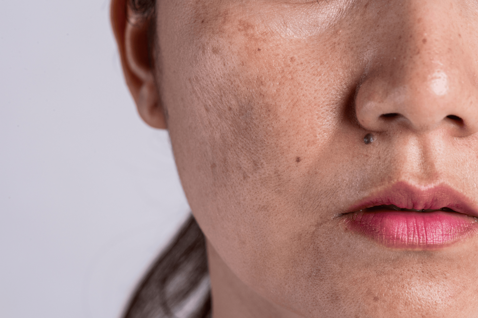
True solar lentigines share consistent features. They are uniformly tan to dark brown with clearly defined, regular borders. The color remains consistent throughout the spot, with no darker areas, lighter patches, or reddish tones. Size typically ranges from a few millimeters to about one centimeter, though they can occasionally grow larger. The surface texture matches the surrounding skin exactly: smooth, flat, and without any scaling or roughness.
Solar lentigines are different from freckles and moles. Freckles are genetic, appear in childhood, and often fade during the winter months. Moles are raised growths that can appear anywhere on the body, regardless of sun exposure. Solar lentigines are always completely flat, never raised, and appear only on sun-exposed areas.
Solar lentigines appear exclusively in areas of the skin that receive chronic sun exposure. The face, particularly the forehead, cheeks, and nose, develops spots first. The backs of hands and forearms follow closely, especially in people who drive frequently with windows down. The upper back, shoulders, and chest are common in those who spend time outdoors.
When a Sun Spot Requires Professional Evaluation
Most solar lentigines never cause problems. But knowing when to seek evaluation prevents dangerous delays in the diagnosis of skin cancer.
Dermatologists use the ABCDE criteria to assess any pigmented lesion. Asymmetry means one half doesn't match the other. Border irregularity shows jagged, notched, or blurred edges. Color variation includes multiple shades of brown, black, red, or blue within the same spot. A diameter larger than 6mm raises concern. Evolution, meaning any change in size, shape, or color, demands immediate attention regardless of the lesion's other features.
Lentigo maligna is a type of melanoma that develops slowly on sun-damaged skin. It looks remarkably similar to early-stage solar lentigo, which makes it particularly dangerous. The key differences: lentigo maligna has irregular borders that seem to fade into surrounding skin, contains multiple color variations, and gradually expands over months or years. It most commonly appears on the faces of older adults with a significant history of sun damage. Melanoma surveillance guidelines note that melanoma can develop in existing spots or on seemingly normal skin, underscoring the importance of monitoring any changes.
Dermatologists generally recommend full-body skin exams every 12 months for adults with risk factors such as fair skin, high UV exposure, or a personal history of skin cancer. A dermatologist can identify suspicious lesions you might dismiss as normal aging, using dermoscopy to see structures invisible to the naked eye. Doctronic.ai provides an accessible first step, offering AI-powered assessments that help determine whether a spot warrants an in-person dermatology appointment.
For more on how brown spots develop and what distinguishes harmless ones from those worth monitoring, see this related overview of brown spots on skin and sun damage.
Treatment Options for Solar Lentigines
While solar lentigines don't require treatment for health reasons, many people choose to have them removed for cosmetic reasons.
Prescription retinoids like tretinoin increase cell turnover, gradually fading solar lentigines over several months. Combination products containing retinoids and depigmenting agents produce faster results than either alone. Over-the-counter options with vitamin C, niacinamide, or azelaic acid offer gentler alternatives, though results take longer. Consistency matters more than product strength, with daily application for three to six months producing visible improvement.
Cryotherapy freezes individual spots with liquid nitrogen, causing pigmented cells to die and slough off within one to two weeks. Chemical peels use acids to remove the outer layers of skin where pigment is concentrated. Both procedures carry slight risks of scarring or temporary darkening, particularly in darker skin tones.
Laser treatments target melanin specifically, breaking up pigment clusters while leaving surrounding tissue intact. Q-switched lasers and picosecond lasers produce excellent results with minimal downtime. Most patients need two to four sessions spaced several weeks apart. Post-treatment sun protection is critical: UV exposure can cause treated areas to darken again.
Prevention Strategies
Preventing new solar lentigines is far easier than treating existing ones.
Daily sunscreen application is non-negotiable, even on cloudy days. Choose SPF 30 or higher with broad-spectrum protection against both UVA and UVB rays. Apply generously and reapply every two hours during outdoor activities. Sun-protective clothing with UPF ratings provides reliable coverage without reapplication concerns. Wide-brimmed hats protect the face and neck, areas particularly prone to solar lentigines.
Topical antioxidants like vitamin C, vitamin E, and ferulic acid neutralize free radicals generated by UV exposure. They don't replace sunscreen but provide an additional layer of defense. Dietary antioxidants from berries, leafy greens, and green tea support skin repair from within.
Frequently Asked Questions
Solar lentigines themselves don't transform into cancer. The concern is that lentigo maligna, an early form of melanoma, can be mistaken for a harmless sunspot. Regular monitoring catches any changes early, which is why dermatologists recommend annual skin exams for anyone with an extensive sun exposure history.
Freckles are genetic, typically appear in childhood, and often fade in winter. Solar lentigines appear in adulthood, result from cumulative UV exposure, don't fade seasonally, and tend to darken with additional sun exposure. They're also usually larger and more sharply defined than freckles.
Yes. "Liver spots" is an outdated term for solar lentigines. They have no connection to liver function; the name came from their brownish color.
Any rapidly appearing brown spot warrants evaluation. True solar lentigines develop gradually over the years. A sudden appearance suggests a different type of lesion that requires professional assessment.
See a dermatologist if a spot has irregular or blurred borders, contains multiple colors, grows larger than a pencil eraser, or changes in any way over weeks to months. When in doubt, get it checked rather than waiting.
Most patients see visible fading within 4 to 8 weeks after treatment, with complete clearance often requiring multiple sessions spaced 4 to 6 weeks apart depending on device type and skin tone.
The Bottom Line
Solar lentigo serves as a visible reminder of cumulative sun damage that demands ongoing attention rather than dismissal. The spots themselves are benign, but their resemblance to lentigo maligna means any change in size, color, or shape warrants prompt evaluation. For convenient monitoring of skin changes and personalized guidance on when to seek professional care, visit Doctronic.ai for AI-powered medical consultations available 24/7.
What Causes Solar LentiginesThat flat brown spot on the back of your hand probably appeared so gradually you barely noticed it. Solar lentigo, commonly called a sun spot or [...]
Join 50,000+ readers using Doctronic to understand symptoms, medications, and next steps.
Only one more step.
Add your phone number below to get health updates and exclusive VIP offers.
By providing your phone number, you agree to receive SMS updates from Company. Message and data rates may apply. Reply “STOP” to opt-out anytime. Read our Privacy Policy and Terms of Service for more details.
Thanks for subscribing
Save your consults. Talk with licensed doctors and manage your health history.