
Differin (Adapalene) for Seniors: What to Know
Read More

Medically reviewed by Oghenefejiro Okifo | MD , Harvard Medical School | Henry Ford Hospital - Detroit, MI on April 25th, 2026. Updated on April 30th, 2026
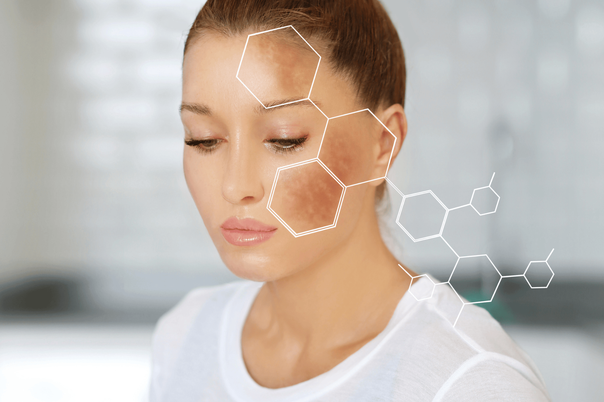
Brown spots on skin develop from melanin overproduction triggered by UV exposure, hormonal changes, or genetics
Solar lentigines, seborrheic keratoses, and freckles are typically harmless and require no medical treatment
The ABCDE checklist helps identify suspicious spots that may indicate melanoma
Actinic keratosis serves as a precancerous warning sign requiring professional evaluation
Monthly self-exams and annual dermatologist visits catch concerning changes early
Noticed a new or changing brown spot? Doctronic.ai offers 24/7 AI-powered consultations to help evaluate your skin concerns
Brown spots on the skin are one of the most common concerns people bring to dermatologists. These marks range from barely noticeable freckles to dark patches that seem to appear overnight. The question everyone asks: are these harmless sun damage or something to watch closely?
The answer depends on several factors. Most brown spots result from years of sun exposure and pose no health threat. Understanding what causes these spots and knowing the warning signs can protect long-term skin health.
Melanin is the pigment responsible for skin, hair, and eye color. When UV rays hit the skin, specialized cells called melanocytes produce extra melanin as a protective response. This pigment absorbs harmful radiation and shields deeper skin layers from damage.
Over time, melanin production can become uneven. Some melanocytes produce more pigment than others, creating concentrated areas of color. This uneven distribution results in the brown spots that become more visible with age.
Sun exposure remains the primary cause of brown spots. Years of UV damage accumulate in the skin, and melanocytes eventually respond by producing permanent patches of excess pigment.
Hormonal changes also trigger hyperpigmentation. Pregnancy, birth control pills, and hormone replacement therapy can cause melasma, a condition producing symmetrical brown patches on the face. These hormonal spots often fade when hormone levels stabilize.
Solar lentigines are flat, tan to dark brown spots that appear on sun-exposed areas. They commonly develop on the face, hands, shoulders, and arms after age 40. These spots have clearly defined borders and uniform color throughout.
True age spots do not require treatment but signify significant sun exposure. Their presence indicates the skin has absorbed substantial UV radiation over the years. While harmless themselves, they serve as reminders to protect skin from further damage.
Seborrheic keratoses look different from flat age spots. These growths appear waxy, raised, and sometimes scaly. They range from light tan to nearly black and often have a "stuck-on" appearance.
These growths are completely benign and extremely common after age 50. They can appear anywhere on the body except the palms and soles. Though sometimes cosmetically bothersome, seborrheic keratoses pose no cancer risk.
Freckles, or ephelides, are small brown spots that appear in childhood and darken with sun exposure. They are most common in people with fair skin and red or blonde hair. Unlike age spots, freckles often fade during the winter months when UV exposure decreases.
Genetic factors determine who develops freckles. The MC1R gene variant makes certain individuals more prone to freckling. These spots are harmless and simply indicate how the skin responds to sunlight.
Dermatologists use the ABCDE system to evaluate suspicious spots. This checklist helps identify characteristics that warrant professional examination:
Asymmetry: one half does not match the other
Border: edges are irregular, ragged, or blurred
Color: multiple shades of brown, black, red, or white within one spot
Diameter: larger than 6mm, about the size of a pencil eraser
Evolving: any change in size, shape, color, or symptoms
Any spot meeting one or more criteria deserves evaluation. Doctronic.ai can help assess initial concerns and recommend whether in-person dermatology visits are needed.
Actinic keratoses are rough, scaly patches caused by sun damage. They feel like sandpaper and may be pink, red, or brown. These lesions develop on sun-exposed areas and indicate significant UV damage.
Unlike harmless age spots, actinic keratoses can progress to squamous cell carcinoma if left untreated. Roughly 5 to 10% of these lesions may progress to invasive squamous cell carcinoma over several years. Early treatment prevents this progression and eliminates the precancerous cells.
The ugly duckling sign refers to a mole or spot that looks different from surrounding lesions. Most people's moles share similar characteristics in color, size, and shape. A spot that stands out from this pattern deserves attention.
This approach helps identify melanomas that might not meet all ABCDE criteria. If one spot looks noticeably different from others, schedule a professional evaluation regardless of its specific characteristics.
Dermoscopy uses a specialized magnifying device to examine skin structures invisible to the naked eye. This tool reveals pigment patterns, blood vessel arrangements, and other features that help distinguish benign spots from concerning lesions.
The examination is painless and takes only minutes. Dermatologists can evaluate multiple spots in a single visit and determine which require further investigation.
Biopsies remove a small tissue sample for laboratory analysis. This procedure provides a definitive diagnosis when visual examination leaves questions. Local anesthesia makes the process comfortable, and results typically return within one to two weeks.
Not every suspicious spot requires a biopsy. Dermatologists weigh clinical findings, patient history, and risk factors when recommending this step.
Prescription retinoids increase cell turnover and gradually fade brown spots. These vitamin A derivatives also improve overall skin texture and reduce fine lines. Results require consistent use over several months.
Vitamin C serums provide antioxidant protection and inhibit melanin production. Applied daily, they can lighten existing spots while preventing new ones from forming. Look for products containing L-ascorbic acid at 10 to 20% concentration or its stabilized derivatives for best results.
Laser treatments target melanin directly, breaking up pigment deposits that the body then clears naturally. Multiple sessions may be needed depending on spot depth and darkness.
Chemical peels remove the outer skin layers, taking superficial pigmentation with them. Cryotherapy freezes individual spots with liquid nitrogen, causing them to blister and peel away. Both options work well for isolated age spots.
Daily sunscreen application prevents new brown spots and protects existing ones from darkening. Choose broad-spectrum formulas with SPF 30 or higher. Reapply every two hours during extended outdoor exposure.
Physical barriers add another protection layer. Wide-brimmed hats shield the face and neck. UV-protective clothing covers arms and shoulders without requiring constant sunscreen reapplication.
Monthly skin checks catch changes early when treatment is most effective. Examine the entire body systematically, including areas that rarely see the sun. Use mirrors to view the back, scalp, and other hard-to-see regions.
Document spots with photographs to track changes over time. Note any new lesions or alterations to existing ones. Bring this information to annual dermatologist appointments for a comprehensive evaluation.
Yes, brown spots can seem to appear overnight, especially after significant sun exposure or hormonal changes. Spots that appear very rapidly or change quickly should be evaluated by a dermatologist.
Freckles may fade during winter months, and some pregnancy-related melasma resolves after delivery. True age spots and seborrheic keratoses are permanent without treatment.
Color alone does not determine danger. Multiple colors within a single spot raise more concern than overall darkness. The ABCDE criteria provide better guidance than color intensity alone.
Most dermatologists recommend a full-body skin examination every 12 months. Individuals with high risk factors may need checks every 6 months.
Brown spots on skin usually represent harmless sun damage, but knowing the warning signs protects against serious conditions. Monthly self-exams and annual professional screenings catch concerning changes when they are most treatable. For quick skin concern evaluations, visit Doctronic.ai for 24/7 AI-powered consultations that help determine your next steps.

Join 50,000+ readers using Doctronic to understand symptoms, medications,
and next steps.
Add your phone number below to get health updates and exclusive VIP offers.
By providing your phone number, you agree to receive SMS updates from Company. Message and data rates may apply. Reply “STOP” to opt-out anytime. Read our Privacy Policy and Terms of Service for more details.
Save your consults. Talk with licensed doctors and manage your health history.