
Red Moles on Skin: Cherry Angiomas and When to Be Concerned
What Are Cherry Angiomas?If you have noticed a small, bright red dot on your skin that seems to have appeared out of nowhere, there is a good chance it is a cherry angioma. [...]
Read More
Medically reviewed by Alan Lucks | MD , Alan Lucks MDPC Private Practice - New York on May 24th, 2026. Updated on May 28th, 2026
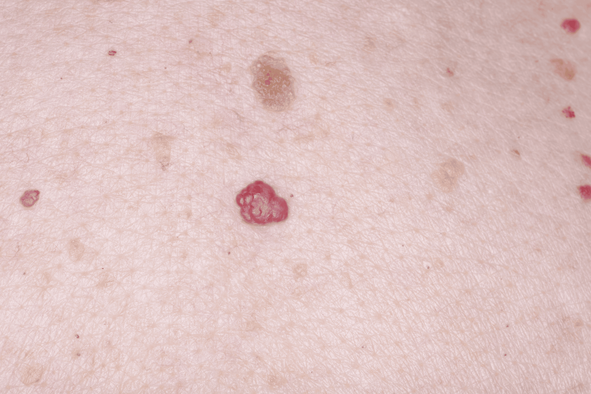
Cherry angiomas are small, bright red growths made of overgrown blood vessels near the skin's surface. They are among the most common benign skin growths in adults.
They can appear anywhere on the body, are typically round or dome-shaped, and range from pinpoint size to about a quarter inch in diameter.
Cherry angiomas are not contagious, not cancerous, and do not require treatment unless they bleed frequently or bother you cosmetically.
A red spot that bleeds easily, changes shape rapidly, has an irregular border, or appears in a child warrants a dermatologist evaluation.
Removal options include laser therapy, electrocautery, and shave excision. All are quick in-office procedures.
Doctronic.ai connects you with a licensed physician who can evaluate an unusual red spot and advise on whether treatment is needed.
If you have noticed a small, bright red dot on your skin that seems to have appeared out of nowhere, there is a good chance it is a cherry angioma. These common growths develop when clusters of small blood vessels (capillaries) dilate and proliferate just beneath the skin's surface, creating a visible red bump.
Cherry angiomas are also called senile angiomas or Campbell de Morgan spots. Despite the name "senile," they are not a sign of decline. They become more frequent with age, and by 70, most adults have at least one. Cherry angiomas are entirely benign vascular growths with no malignant potential. For guidance on how to tell cherry angiomas apart from other skin spots, the guide about moles vs. birthmarks covers common benign skin growths and the signs that distinguish them from lesions worth evaluating.
Cherry angiomas are typically:
Bright red, cherry red, or occasionally purple-red in color
Round or slightly dome-shaped
Smooth-surfaced, sometimes slightly raised
Between 1 and 5 millimeters in diameter, though larger ones exist
Painless unless irritated
They can appear anywhere on the body but are most common on the trunk (chest, back, and abdomen), arms, and shoulders. They are rarely found on the face, palms, or soles.
When pressed with a finger, a cherry angioma typically blanches (turns lighter or white) because the blood is pushed out of the dilated vessels. This blanching response distinguishes it from petechiae (small pinpoint bruises that do not blanch) and helps confirm its vascular origin.
Cherry angiomas affect people of all skin tones and ethnicities. Several factors are associated with their development:
Age is the strongest predictor. They are uncommon before age 30 and become progressively more frequent afterward.
Hormonal changes, particularly during pregnancy, can trigger new cherry angiomas or cause existing ones to enlarge temporarily.
Some research suggests a genetic component. If a parent developed many cherry angiomas, you may be more likely to as well.
Certain chemical exposures, including bromides and mustard gas, have been associated with widespread eruptions of cherry angiomas, though this is uncommon in everyday life.
Cherry angiomas are not caused by sun damage, diet, or poor health. Developing them does not signal any underlying disease.
Most cherry angiomas require no evaluation beyond recognizing them. However, a few situations warrant a closer look:
A red spot that bleeds repeatedly or bleeds heavily from minor contact should be examined. Cherry angiomas can bleed if scratched or rubbed, but persistent or heavy bleeding is worth discussing with a doctor.
Rapid changes in size or shape are not typical of cherry angiomas. If a red lesion grows quickly, develops irregular borders, or changes color significantly over a short period, have it evaluated.
Angiokeratomas are another type of vascular lesion that can resemble cherry angiomas. They tend to be darker red or purple-black, have a rougher or keratotic (scaly) surface, and can occur on the genitalia or lower extremities. Some types of angiokeratomas are associated with underlying systemic conditions, so an accurate diagnosis is important.
A lesion that does not blanch on compression, is growing on a child, or is accompanied by other unexplained symptoms also merits professional evaluation.
When in doubt, a dermatologist can examine the spot with a dermatoscope to confirm the diagnosis. If the appearance is ambiguous, a biopsy provides a definitive answer.
Cherry angiomas do not require treatment. Many people choose removal for cosmetic reasons or because a spot catches on clothing and bleeds. Common in-office methods include:
Laser therapy uses a pulsed-dye or Nd:YAG laser to target the blood vessels within the angioma. The vessel absorbs the laser energy, collapses, and is reabsorbed by the body over one to two weeks. Most cherry angiomas resolve in a single session.
Electrocautery applies a low electrical current to cauterize the blood vessels. The treated tissue is destroyed and sloughs off over several days. This method is fast and well-suited for flat or slightly raised lesions.
Shave excision removes the growth with a thin surgical blade at the level of the surrounding skin. No deep incision is needed, and the area heals within one to two weeks.
Cryotherapy freezes the lesion with liquid nitrogen, destroying the tissue. It is effective but may require more than one treatment session for larger angiomas.
All procedures are performed under local anesthesia. Recovery is brief, and scarring is uncommon when performed by a trained provider.
No. A blood blister forms when trauma causes blood to pool between layers of skin, creating a fluid-filled blister that appears dark red or purple. It is painful and resolves on its own as the blood is absorbed. A cherry angioma is a permanent vascular growth made of overgrown capillaries. It is painless, does not fluctuate in size, and does not resolve without treatment.
Occasionally, a very small cherry angioma may fade or disappear, but the vast majority are permanent unless treated. New ones tend to develop over time as a person ages.
No. Home removal methods (including cutting, scraping, or applying unregulated topical products) risk infection, permanent scarring, and excessive bleeding. More importantly, removing a lesion at home eliminates the possibility of a professional diagnosis. If you are certain the spot is a cherry angioma and want it removed, see a dermatologist.
Yes. Some people develop dozens, or even hundreds, over their lifetimes. A sudden eruption of many new cherry angiomas over a short period is less typical and may occasionally be associated with hormonal changes or, rarely, an underlying condition. A dermatologist can evaluate whether further workup is appropriate.
They can bleed if scratched, shaved over, or subjected to friction. The bleeding typically stops quickly with mild pressure. If a cherry angioma bleeds frequently or bleeds heavily, that is a reason to have it evaluated and potentially removed.
Cherry angiomas are benign vascular growths and do not become cancerous. If a red lesion is changing, growing irregularly, or does not match the typical appearance of a cherry angioma, it should be evaluated to rule out other causes, including rare vascular tumors or amelanotic (non-pigmented) melanoma.
Cherry angiomas are common and harmless, needing treatment only if they bleed repeatedly or bother you cosmetically. A red mole that changes quickly, bleeds heavily, or appears in a child is worth having examined — a dermatologist can give you a clear diagnosis in one visit.
If you are unsure about a spot, Doctronic.ai lets you connect with a licensed physician in a real-time online visit.
What Are Cherry Angiomas?If you have noticed a small, bright red dot on your skin that seems to have appeared out of nowhere, there is a good chance it is a cherry angioma. [...]
Read More
Join 50,000+ readers using Doctronic to understand symptoms, medications,
and next steps.
Add your phone number below to get health updates and exclusive VIP offers.
By providing your phone number, you agree to receive SMS updates from Company. Message and data rates may apply. Reply “STOP” to opt-out anytime. Read our Privacy Policy and Terms of Service for more details.
Save your consults. Talk with licensed doctors and manage your health history.