Diseases and Conditions
Understanding Milia: Small White Bumps on Your Skin
February 5, 2026
2 min read

Medically reviewed by Alan Lucks | MD, Alan Lucks MDPC Private Practice - New York on May 23rd, 2026.
Published on April 15th, 2026. Updated on September 11th, 2026.
White spots on skin result from various conditions ranging from harmless fungal infections to autoimmune disorders like vitiligo.
Tinea versicolor, caused by yeast overgrowth, is one of the most common and treatable causes of skin discoloration.
Between 0.76% and 1.11% of adults in the United States have vitiligo, making it more common than many people realize.
Texture changes, itching, or rapid spreading of white spots warrant a visit to a healthcare provider.
Treatment options include antifungal creams, corticosteroids, light therapy, and cosmetic camouflage techniques.
Doctronic.ai offers free AI doctor consultations to help identify potential causes before scheduling an in-person appointment.
Noticing white spots on your skin can feel alarming. Most people immediately worry about serious conditions, but the reality is far more reassuring. The majority of white skin patches are benign and respond well to treatment. Understanding what causes these spots and knowing when they require medical attention helps separate genuine concerns from minor cosmetic issues.
Hypopigmentation refers to any loss of skin color and occurs when melanocytes (the cells responsible for producing pigment) are damaged, destroyed, or simply stop functioning properly. The skin contains billions of these specialized cells, and disruptions in their function appear as lighter patches against the surrounding tissue. Some causes are temporary and resolve on their own. Others require ongoing management. Knowing the difference matters for peace of mind and proper care.
Melanin gives skin its color. When production drops in certain areas, white or light-colored patches appear. This process can happen for several reasons: fungal infections, autoimmune responses, genetic factors, sun damage, or inflammatory skin conditions. Each cause produces slightly different patterns and textures.
Environmental factors play a significant role. Excessive sun exposure damages melanocytes over time, while certain medications can interfere with pigment production. Hormonal changes during pregnancy or thyroid dysfunction may also trigger hypopigmentation in some individuals. The good news is that identifying the underlying cause usually points directly to effective treatment options. Doctronic.ai can help assess symptoms and suggest whether professional evaluation is needed.
A yeast called Malassezia naturally lives on everyone's skin. When it overgrows (often due to heat, humidity, or oily skin), it interferes with normal pigmentation. The result is tinea versicolor: patches of lighter or darker skin, typically on the chest, back, and upper arms.
These spots often become more noticeable after sun exposure because affected areas do not tan like surrounding skin. The condition is not contagious and responds quickly to antifungal treatments. Many people experience recurring episodes, especially during warm months.
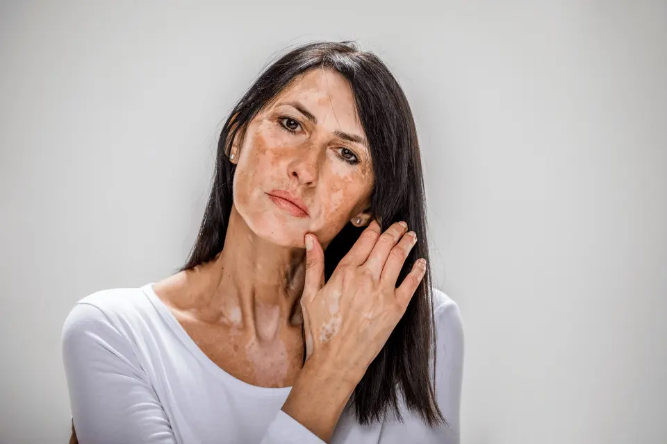
Vitiligo occurs when the immune system mistakenly attacks melanocytes, destroying them completely in certain areas. This creates stark white patches that can appear anywhere on the body. Research indicates that between 0.76% and 1.11% of adults in the United States have this condition.
The patches often appear symmetrically on both sides of the body. Vitiligo can begin at any age, but it frequently starts before age 30. While there is no cure, treatments can help restore some pigmentation or slow progression.
Children and teenagers commonly develop pityriasis alba, a mild form of eczema that leaves pale, slightly scaly patches on the face, arms, and torso. The condition typically appears after a bout of dry skin or mild eczema and is more visible on darker skin tones.
These patches are harmless and usually fade on their own over months or years. Keeping skin moisturized helps speed recovery. No aggressive treatment is typically necessary.
Small, flat white spots that appear on sun-exposed areas such as the arms and legs often indicate idiopathic guttate hypomelanosis. These spots develop from cumulative sun damage over decades and become more common after age 40. Unlike age spots, which darken with UV exposure, these patches are permanently depigmented areas resulting from long-term damage to melanocytes.
The spots are permanent but purely cosmetic. They do not spread, itch, or indicate any underlying health problem. Sun protection can prevent new spots from forming.
Pay attention to how white spots feel, not just how they look. Smooth, flat patches that do not itch usually indicate benign conditions. Raised, scaly, or itchy spots may suggest fungal infections or inflammatory conditions requiring treatment.
Any new or changing spots on the skin should be evaluated by a healthcare provider. This is especially true for spots with irregular borders, multiple colors, or those that bleed easily.
Slow-developing spots over the years rarely signal urgent problems. Rapid appearance of new patches within weeks or months warrants prompt evaluation. Similarly, white spots accompanied by hair turning white in the same area may indicate progression of vitiligo.
Symmetric distribution (matching patches on both arms or both legs) often points toward autoimmune causes rather than fungal infections. Documenting when spots first appeared and how they have changed helps healthcare providers make accurate diagnoses.
Dermatologists use several tools to identify the cause of white spots. A Wood's lamp examination uses ultraviolet light to reveal fungal infections that glow specific colors. Skin scrapings examined under a microscope can confirm tinea versicolor within minutes.
For suspected vitiligo, doctors may order blood tests to check thyroid function and screen for other autoimmune conditions. Skin biopsies are occasionally necessary when the diagnosis remains unclear. Doctronic.ai provides initial guidance on which symptoms suggest specific conditions, helping users prepare for medical appointments with relevant questions.
Tinea versicolor typically clears with over-the-counter antifungal creams containing ketoconazole or selenium sulfide. Applying these products to affected areas for two to four weeks usually eliminates the fungal overgrowth. Prescription-strength options exist for stubborn cases.
Mild corticosteroid creams help reduce inflammation in conditions like pityriasis alba. These should be used sparingly on the face and only as directed by a healthcare provider to avoid skin thinning. Oral steroids or immunosuppressants are sometimes used.
This is an FDA-approved treatment for the underlying inflammation.
Narrowband UVB phototherapy represents the gold standard for treating vitiligo. Regular sessions over several months can stimulate melanocytes to repopulate affected areas. Success rates vary depending on the location and extent of pigment loss.
Excimer laser treatments offer targeted therapy for smaller vitiligo patches. These concentrated light sessions require fewer treatments than full-body phototherapy booths.
Specialized makeup products designed for vitiligo provide immediate cosmetic improvement. Waterproof formulations can last through swimming and sweating. Self-tanning products also help blend white patches with the surrounding skin.
Sun protection is essential for all hypopigmentation conditions. Unpigmented skin burns easily and lacks natural UV defense. Daily broad-spectrum sunscreen with SPF 30 or higher protects vulnerable areas and helps prevent new sunspots from forming.
Preventing white spots depends on their cause. For fungal-related discoloration, keeping the skin clean and dry helps reduce yeast overgrowth. Wearing breathable fabrics during hot weather helps maintain healthy skin balance.
Sun protection prevents idiopathic guttate hypomelanosis and protects existing vitiligo patches from sunburn. Establishing a consistent moisturizing routine supports overall skin barrier function and may reduce episodes of pityriasis alba in children.
Regular skin self-examinations help catch changes early. Photographing existing spots creates a baseline for tracking any progression over time.
Most causes of white spots are not contagious. Vitiligo, pityriasis alba, and sun damage spots cannot spread through contact. Tinea versicolor involves a yeast that naturally lives on everyone’s skin, so direct transmission is extremely unlikely.
Tinea versicolor and pityriasis alba typically resolve fully with treatment or time, though repigmentation may take several months after the underlying condition clears. Vitiligo patches may partially repigment with treatment but often require ongoing management.
White spots are rarely associated with skin cancer. Melanoma typically appears as dark, irregular spots rather than light ones. Pale patches that do not heal or change significantly should still be evaluated to rule out rare conditions.
Children with white facial patches likely have pityriasis alba, which is harmless. Consultation becomes important if spots spread rapidly, appear on unusual body areas, or are accompanied by other symptoms such as changes in hair color.
White spots on the skin usually result from treatable conditions like fungal infections or manageable autoimmune responses. Understanding the cause helps determine whether home treatment suffices or professional care is needed. For quick guidance on skin concerns, visit Doctronic.ai for free AI consultations that help identify potential causes and recommend next steps.
Get advice about white spots on skin
Chat Now
Join 50,000+ readers using Doctronic to understand symptoms, medications,
and next steps.
Add your phone number below to get health updates and exclusive VIP offers.
By providing your phone number, you agree to receive SMS updates from Company. Message and data rates may apply. Reply “STOP” to opt-out anytime. Read our Privacy Policy and Terms of Service for more details.
Save your consults. Talk with licensed doctors and manage your health history.