A dysplastic nevus is an atypical mole with irregular features that may increase melanoma risk and requires professional evaluation.
The ABCDE criteria (Asymmetry, Border, Color, Diameter, Evolution) help identify moles needing further assessment.
Approximately 2 to 8% of the White population in the United States has one or more atypical nevi.
Regular skin self-exams and the "ugly duckling" sign are essential tools for early detection.
Management ranges from clinical observation to surgical excision depending on severity grading.
Doctronic.ai offers AI-powered consultations to help evaluate skin concerns and determine when a dermatology appointment is needed.
What Everyone Should Know About Atypical Moles
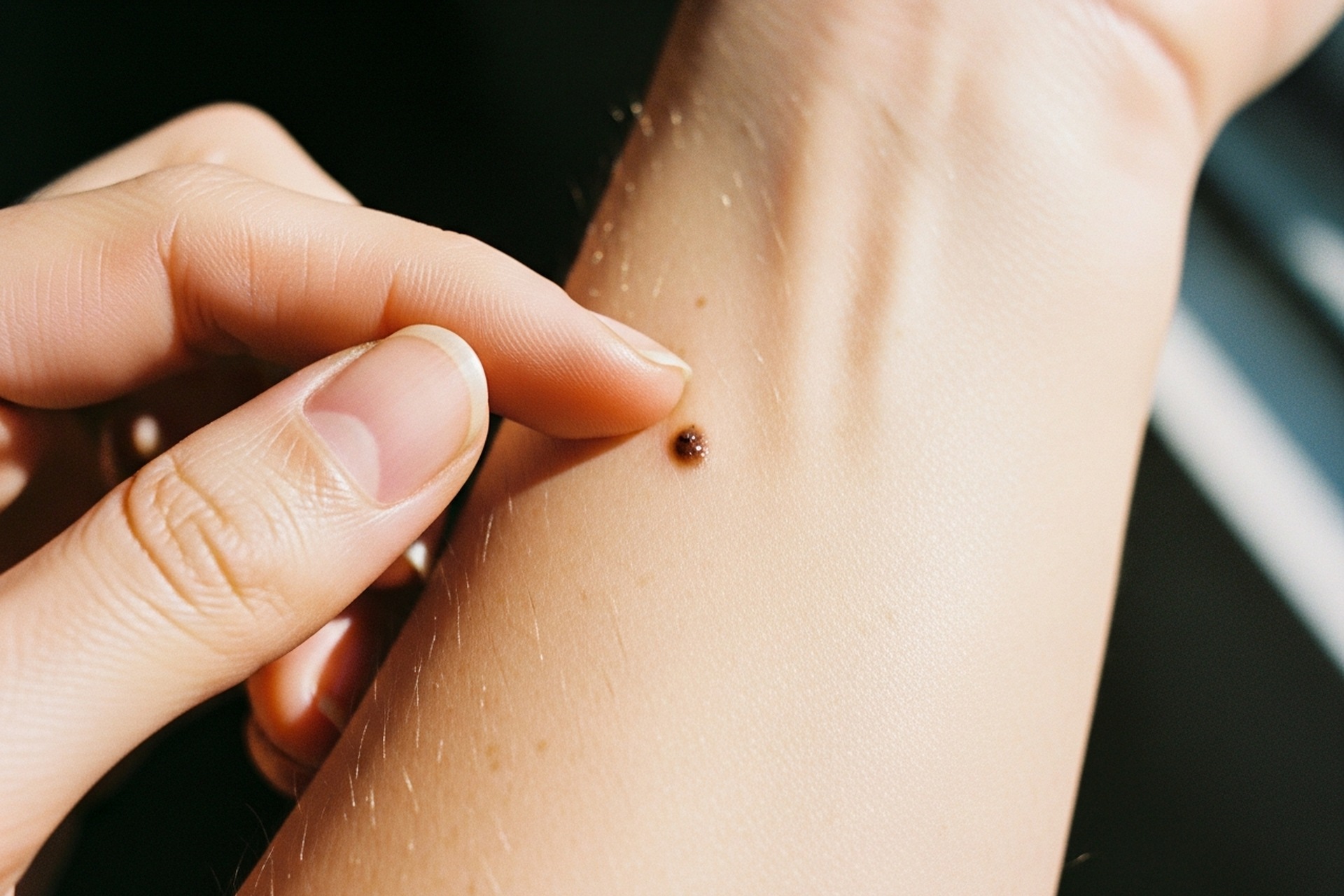
That odd-looking mole on your arm might be nothing to worry about, or it might deserve a closer look. A dysplastic nevus is an atypical mole that looks different from common moles in shape, color, or size.
These moles are not cancer, but they can signal a higher melanoma risk. With approximately 100,600 new cases of invasive melanoma expected in the United States in 2026, understanding when an atypical mole needs further evaluation could save your life.
Understanding Dysplastic Nevi
Dysplastic nevi sit somewhere between normal moles and melanoma on the spectrum of skin growths. They often appear during adolescence or young adulthood and can continue developing throughout life.
The ABCDEs of Atypical Moles
Dermatologists use the ABCDE system to evaluate suspicious moles:
Asymmetry: One half does not match the other half.
Border irregularity: Ragged, notched, or blurred edges rather than smooth, defined margins.
Color variation: Multiple shades of brown, tan, black, red, or blue within a single mole.
Diameter: Exceeds 6 millimeters, roughly the size of a pencil eraser.
Evolution: Any change in size, shape, color, or symptoms like itching or bleeding over time.
How Dysplastic Nevi Differ from Common Moles
Common moles are usually smaller than 5 millimeters with even coloring and distinct borders. Dysplastic nevi tend to be larger, with fuzzy or fading edges that blend into surrounding skin.
The surface texture may appear pebbly or slightly raised in the center with a flat edge. Color patterns are typically uneven, mixing different shades of brown with pink or red areas. These moles can appear anywhere, but frequently develop on sun-exposed areas like the back, chest, and legs.
Risk Factors and the Link to Melanoma
Having dysplastic nevi does not mean someone will develop melanoma. These moles do indicate increased risk that warrants attention and monitoring.
The Role of Genetics and Family History
Family history plays a significant role in dysplastic nevus development. People with close relatives who have had melanoma or multiple atypical moles face a higher risk.
Familial atypical multiple mole melanoma syndrome, a genetic condition, causes numerous dysplastic nevi and substantially increases melanoma likelihood. An estimated 2 to 8% of the White population has one or more atypical nevi, making this a relatively common finding that still requires appropriate surveillance.
Cumulative Sun Exposure and Skin Type
People with fair skin, light hair, and light eyes face a higher risk for both dysplastic nevi and melanoma. Cumulative sun exposure throughout life, especially sunburns during childhood, contributes to atypical mole development.
Indoor tanning significantly increases risk regardless of skin type. Those who work outdoors or live in sunny climates should be particularly vigilant about monitoring their skin.
When to Seek Professional Evaluation
Knowing when to see a dermatologist can feel confusing. Not every unusual mole requires immediate medical attention, but certain signs should prompt a visit. Doctronic.ai can help assess initial concerns and guide decisions about when an in-person evaluation is necessary.
The Ugly Duckling Sign
The ugly duckling sign is one of the most useful tools for spotting concerning moles. Most moles on a person's body tend to look similar to each other. When one mole looks noticeably different from the rest, it stands out like an ugly duckling among its siblings.
This mole deserves extra attention even if it does not meet all the ABCDE criteria. The concept works because melanoma often looks different from a person's typical mole pattern. Understanding how to assess skin symptoms builds the visual awareness needed for effective mole monitoring.
Monitoring Changes Over Time
Any mole that changes over weeks or months needs evaluation. Taking photographs of atypical moles helps track subtle changes that might otherwise go unnoticed. New symptoms like itching, tenderness, or bleeding warrant prompt attention.
Diagnostic Procedures and Grading
When a dermatologist examines a suspicious mole, several diagnostic tools help determine whether removal is necessary.
Dermoscopy and Clinical Examination
Dermoscopy uses a specialized magnifying device with polarized light to examine mole structures invisible to the naked eye. This tool reveals pigment patterns, blood vessel arrangements, and other features that help distinguish benign moles from potentially dangerous ones.
A thorough clinical examination includes checking the entire body for additional atypical moles and documenting their locations. Patients who want to understand what a dermatologist looks for can explore telehealth dermatology for an initial assessment before scheduling an in-person visit.
Skin Biopsy and What to Expect
When a mole appears suspicious, the dermatologist may recommend a skin biopsy. This procedure removes all or part of the mole for laboratory examination:
Excisional biopsy: Removes the entire mole with a margin of normal skin.
Shave biopsy: Removes the raised portion of the mole.
Punch biopsy: Takes a deeper, circular sample for analysis.
A pathologist studies the tissue under a microscope to determine whether cells are normal, atypical, or cancerous.
Understanding Atypia Grades
Pathologists grade dysplastic nevi based on how abnormal the cells appear:
Mild atypia: Cells look slightly different from normal. Typically requires monitoring only.
Moderate atypia: More significant changes that may require additional monitoring or re-excision.
Severe atypia: Cellular changes closer to melanoma. Usually requires complete surgical removal with clear margins.
Management and Prevention
Surgical Excision vs. Observation
Mildly atypical moles with clear biopsy margins often require only regular monitoring. Moderately atypical moles may need re-excision to ensure complete removal, especially if margins are unclear. Severely atypical moles typically require surgical excision with wider margins.
Sun Safety and Self-Exams
Prevention starts with consistent habits:
Use broad-spectrum sunscreen with SPF 30 or higher daily.
Wear protective clothing and avoid peak sun hours (10 AM to 4 PM).
Avoid tanning beds entirely.
Perform monthly skin self-exams using a full-length mirror and a hand mirror.
Check between toes, on the scalp, and in other often-overlooked spots.
Take smartphone photos of concerning moles to track changes between appointments.
Doctronic.ai provides 24/7 access to medical guidance for patients, determining when skin changes warrant an urgent appointment versus routine monitoring.
Frequently Asked Questions
A dysplastic nevus can potentially develop into melanoma, though most do not. The risk increases with the number of atypical moles present and severity of atypia. Regular monitoring catches any concerning changes early.
People with multiple dysplastic nevi should typically see a dermatologist every 6 to 12 months for full-body skin examinations. Those with personal or family history of melanoma may need more frequent visits.
Genetic factors influence dysplastic nevi development. People with family members who have atypical moles or melanoma face a higher risk. Genetic counseling may benefit families with multiple affected members.
Not all dysplastic nevi require removal. Mildly atypical moles with clear margins can often be monitored. Removal decisions depend on atypia grade, margin status, and individual risk factors.
The Bottom Line
Understanding when a dysplastic nevus needs further evaluation empowers you to protect your skin health through early detection and appropriate monitoring. The ABCDE criteria, ugly duckling sign, and regular self-exams provide practical tools for catching changes early.
For guidance on skin concerns or questions about atypical moles, visit Doctronic.ai for AI-powered consultations that help determine your next steps toward professional care.
What Everyone Should Know About Atypical MolesThat odd-looking mole on your arm might be nothing to worry about, or it might deserve a closer look. A dysplastic nevus is an [...]
Join 50,000+ readers using Doctronic to understand symptoms, medications, and next steps.
Only one more step.
Add your phone number below to get health updates and exclusive VIP offers.
By providing your phone number, you agree to receive SMS updates from Company. Message and data rates may apply. Reply “STOP” to opt-out anytime. Read our Privacy Policy and Terms of Service for more details.
Thanks for subscribing
Save your consults. Talk with licensed doctors and manage your health history.