
Natural Alternatives to Vibramycin (Doxycycline)
Read More

Medically reviewed by Veronica Hackethal | MD, MSc , Harvard University | University of Oxford | Columbia Vagelos College of Physicians and Surgeons on May 25th, 2026. Published on April 17th, 2026. Updated on July 23rd, 2026.
Both rosacea and the lupus butterfly rash produce redness on the cheeks and nose, but they have distinct characteristics that dermatologists use to differentiate them clinically
The lupus malar rash characteristically spares the nasolabial folds (the lines running from the nose to the corners of the mouth), while rosacea often involves them
Lupus is a systemic autoimmune disease; its facial rash is typically accompanied by other systemic symptoms including joint pain, fatigue, fever, and photosensitivity, whereas rosacea is a skin condition without systemic involvement
Rosacea involves visible blood vessels, papules, and pustules that are not features of the lupus malar rash
Distinguishing the two requires clinical evaluation and, often, blood testing for lupus-specific markers including antinuclear antibodies (ANA)
To connect with a licensed physician who can evaluate facial rashes and recommend appropriate testing, Doctronic.ai offers free AI consultations and affordable telehealth visits available any time
Rosacea and the butterfly rash of lupus are two of the most commonly confused conditions in dermatology, partly because both produce symmetric facial redness across the cheeks and nose, and partly because both are often initially dismissed as sun damage, flushing, or sensitive skin. The distinction matters significantly because the treatment, monitoring requirements, and health implications are entirely different.
Rosacea is a chronic skin condition managed with topical and oral treatments, lifestyle modifications, and sometimes laser therapy. Systemic lupus erythematosus (SLE) is an autoimmune disease that can affect the kidneys, joints, heart, lungs, and nervous system, and it requires ongoing systemic management including immunosuppressive medications. Treating one as the other either fails to control the skin condition or, more seriously, misses an underlying systemic disease that warrants close monitoring.
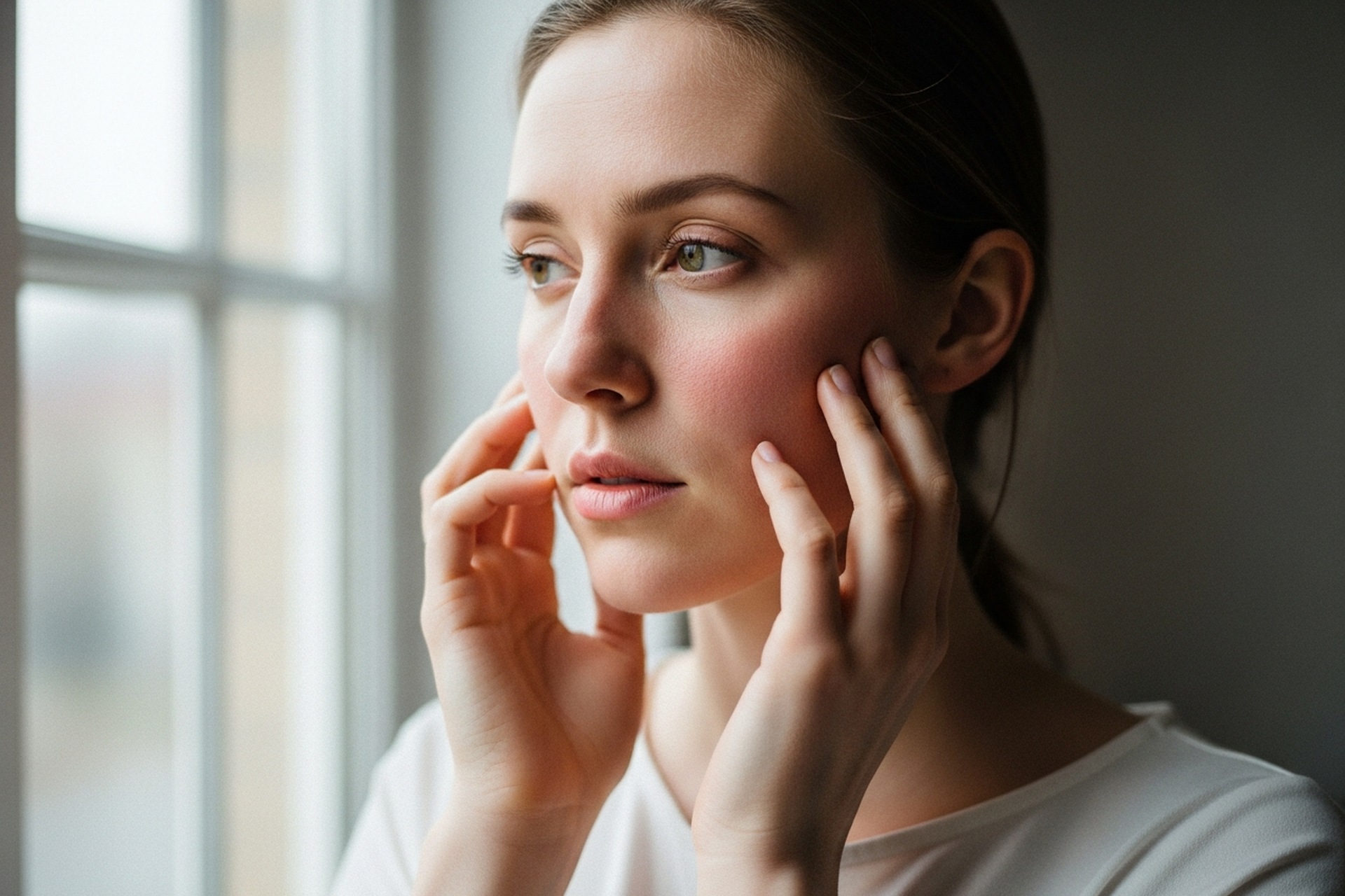
Rosacea is a chronic inflammatory skin condition that primarily affects the central face: the cheeks, nose, chin, and forehead. The hallmark features include persistent redness (erythema), flushing episodes triggered by heat, sun, alcohol, and spicy foods, visible small blood vessels (telangiectasias), and in some subtypes, papules and pustules that resemble acne.
Rosacea commonly involves the nasolabial folds and does not spare them. In phymatous rosacea, the skin thickens, most characteristically on the nose (rhinophyma). Ocular rosacea affects the eyes in about half of rosacea cases, producing symptoms such as eye redness, dryness, and sensitivity to light. Rosacea presents across four subtypes with overlapping but distinct features, and management varies considerably depending on which subtype predominates.
The malar rash of lupus (also called the butterfly rash due to its shape) is a red or pinkish rash that extends across both cheeks and the bridge of the nose in a pattern that visually resembles butterfly wings. The rash is typically flat or slightly raised and can vary in intensity from faint pinkness to deep red or even purple.
The single most clinically useful distinguishing feature of the lupus malar rash is that it characteristically spares the nasolabial folds. The rash does not extend into the natural creases between the nose and mouth, creating a visible border that is often absent in rosacea. This sparing pattern reflects the underlying mechanism: the lupus rash is related to photosensitivity and immune complex deposition rather than vascular reactivity.
The lupus rash is often precipitated or worsened by sun exposure, tends to persist longer than a typical rosacea flush, and does not involve the papules or pustules characteristic of rosacea subtypes. Telangiectasias are not a feature.
Several characteristics reliably help distinguish the two conditions:
Nasolabial fold involvement: Rosacea typically involves them; lupus rash typically spares them. This is the most reliable clinical differentiator.
Papules and pustules: Present in certain rosacea subtypes; absent in lupus malar rash.
Telangiectasias: Common in rosacea; not characteristic of lupus malar rash.
Trigger pattern: Rosacea flushes with heat, exercise, alcohol, spicy food, and emotional stress. The lupus rash worsens with UV exposure specifically.
Systemic symptoms: Rosacea has none. Lupus is associated with joint pain and swelling, persistent fatigue, oral ulcers, fever, photosensitivity, and potential organ involvement.
Duration and behavior: Rosacea persists chronically and flares with identifiable triggers. The lupus malar rash can wax and wane with disease activity.
Accurate identification matters when redness spans the cheeks and nose, a point explored further in a look at facial redness causes.
The butterfly rash is one of the diagnostic criteria for SLE but is present in only about 50 percent of lupus cases, meaning its absence does not rule out lupus. When it is present, it appears alongside the range of systemic lupus symptoms that distinguish lupus from a pure skin condition: joint pain and swelling, fatigue that is often severe and disproportionate to activity level, fever without infection, hair loss, oral ulcers, and photosensitivity.
Lupus can cause kidney inflammation (lupus nephritis), which may produce no symptoms early on but can lead to kidney failure if undetected. Cardiac and pulmonary involvement also occur. This is why identifying lupus accurately and early is clinically significant beyond the skin presentation.
Rosacea is typically diagnosed clinically, based on the appearance of the rash, its distribution, its association with classic triggers, and the absence of systemic findings. Skin biopsy is occasionally performed for atypical presentations.
Lupus diagnosis involves a combination of clinical criteria and blood testing. The antinuclear antibody (ANA) test is the initial screening test; a positive ANA with the right clinical picture warrants further workup including anti-double-stranded DNA antibodies (anti-dsDNA), complement levels, and a complete metabolic panel. A skin biopsy of the malar rash can show immune complex deposits that support the lupus diagnosis.
The ANA test can be positive in people without lupus, so a positive result alone does not diagnose the condition. A negative ANA, however, makes SLE significantly less likely.
Any persistent facial rash that has not been formally diagnosed warrants evaluation. This is particularly true when the rash is accompanied by joint pain, fatigue, oral ulcers, or sun sensitivity, which collectively suggest systemic disease rather than a skin condition. Early lupus diagnosis allows for monitoring and treatment before organ damage occurs.
People already diagnosed with rosacea who develop new systemic symptoms should also seek evaluation, as both conditions can coexist, and rosacea does not exclude the possibility of an independent underlying autoimmune condition.
In their early or mild forms, they can look very similar, which is why laboratory testing and clinical evaluation are important for correct diagnosis. The key visual differentiator is the nasolabial fold: rosacea typically involves it, and the lupus malar rash typically spares it. Papules and telangiectasias in the affected area are more consistent with rosacea.
Not necessarily. ANA tests are positive in many people without lupus, including people with other autoimmune conditions, healthy individuals, and those on certain medications. A positive ANA warrants further evaluation, but lupus diagnosis requires meeting additional criteria including specific antibodies, clinical features, and in some cases biopsy.
Yes. Photosensitivity is a feature of both, though the mechanism is different. Rosacea flushes with sun exposure because UV light triggers vascular reactivity. The lupus malar rash is driven by photosensitivity related to immune activation. Sun protection is relevant in both conditions, though it is particularly important in lupus because sun exposure can trigger systemic flares beyond the skin.
No. The malar butterfly rash appears in approximately 50 percent of people with lupus at some point in the disease course and may not be present when lupus is active in other ways. Lupus can be present and causing internal organ effects without any skin rash at all.
Rosacea is treated with topical agents (metronidazole, azelaic acid, ivermectin), oral antibiotics for inflammatory subtypes, laser treatment for telangiectasias, and trigger avoidance. Lupus treatment is systemic: hydroxychloroquine is the foundational treatment, with corticosteroids and immunosuppressive medications added for more active disease. The two treatments do not overlap significantly.
Rosacea and the lupus butterfly rash are two distinct conditions that share superficial similarities in facial distribution but differ in their underlying causes, clinical features, systemic implications, and treatments. The most reliable clinical differentiator is the nasolabial fold: rosacea involves it, lupus malar rash spares it. Papules, telangiectasias, and classic flush triggers favor rosacea. Joint pain, fatigue, oral ulcers, and photosensitivity are consistent with lupus. Diagnosis requires clinical evaluation and blood testing to screen for lupus-specific markers. Both conditions are manageable with the right treatment, and early accurate diagnosis prevents years of inadequate care. For evaluation of facial rashes by a licensed physician, Doctronic.ai offers affordable telehealth visits available at any time.

Join 50,000+ readers using Doctronic to understand symptoms, medications,
and next steps.
Add your phone number below to get health updates and exclusive VIP offers.
By providing your phone number, you agree to receive SMS updates from Company. Message and data rates may apply. Reply “STOP” to opt-out anytime. Read our Privacy Policy and Terms of Service for more details.
Save your consults. Talk with licensed doctors and manage your health history.