
Can You Take Accutane (Isotretinoin) While Breastfeeding?
Understanding Accutane and Breastfeeding SafetyAccutane, also known by its generic name isotretinoin, is a powerful oral medication used to treat severe acne that hasn't [...]
Read More
Medically reviewed by Lauren Okafor | MD , The Frank H Netter MD School of Medicine, Loyola University Medical Center on April 20th, 2026. Updated on May 28th, 2026
Nodular melanoma accounts for 10-15% of melanoma cases but is responsible for nearly 40% of melanoma deaths because it grows faster and deeper than other types.
Unlike most melanomas, nodular melanoma skips the horizontal spreading phase and immediately invades the deeper layers of skin, making early detection much harder.
The EFG rule helps identify nodular melanoma: Elevation (a new raised bump), Firmness (hard to the touch), and Growth (visibly larger within weeks to a couple of months).
Nodular melanoma can be amelanotic, meaning it lacks the dark pigmentation typically associated with melanoma, making it easy to mistake for a cyst or benign growth.
Breslow thickness is the most important factor in predicting outcomes: tumors under 1mm carry an excellent prognosis, while those over 4mm are associated with significantly worse survival rates.
Doctronic.ai can help you assess a suspicious skin lesion and connect you with a dermatologist before a concerning spot becomes a serious diagnosis.
Most skin cancers, including the more common forms of melanoma, develop gradually. They spread outward across the surface of the skin before they begin growing downward. That horizontal phase gives patients time to notice changes and seek care. Nodular melanoma does not follow that pattern.
This subtype grows vertically from the start. Rather than spreading across the skin, it dives directly into the dermis and subcutaneous tissue. Because it bypasses the surface-spreading phase entirely, it can reach dangerous depths before it becomes visually obvious. By the time many patients notice something wrong, the tumor has already grown considerably thicker.
This behavior explains why nodular melanoma is disproportionately lethal. Although it represents only 10-15% of all melanoma diagnoses, it accounts for approximately 40% of melanoma-related deaths. The timeline from first appearance to a life-threatening stage can be measured in weeks or a few months, not years.
The traditional ABCDE checklist (Asymmetry, Border, Color, Diameter, Evolving) was designed primarily for detecting superficial spreading melanoma. While it remains valuable, nodular melanoma often does not fit those criteria because it does not spread horizontally. A different set of warning signs applies here.
Dermatologists use the EFG rule specifically for nodular melanoma:
Elevation refers to a new raised bump on the skin. Unlike a flat mole that develops slowly over years, this type of lesion appears as a dome-shaped nodule that visibly protrudes from the skin surface.
Firmness means the bump feels hard, similar to the texture of a marble or firm lump under the skin. This density reflects the rapid proliferation of abnormal cells at depth.
Growth describes how quickly the lesion changes. Nodular melanoma can double in size within weeks to a couple of months. If you notice a bump growing at that pace, it should be evaluated immediately regardless of its color or appearance.
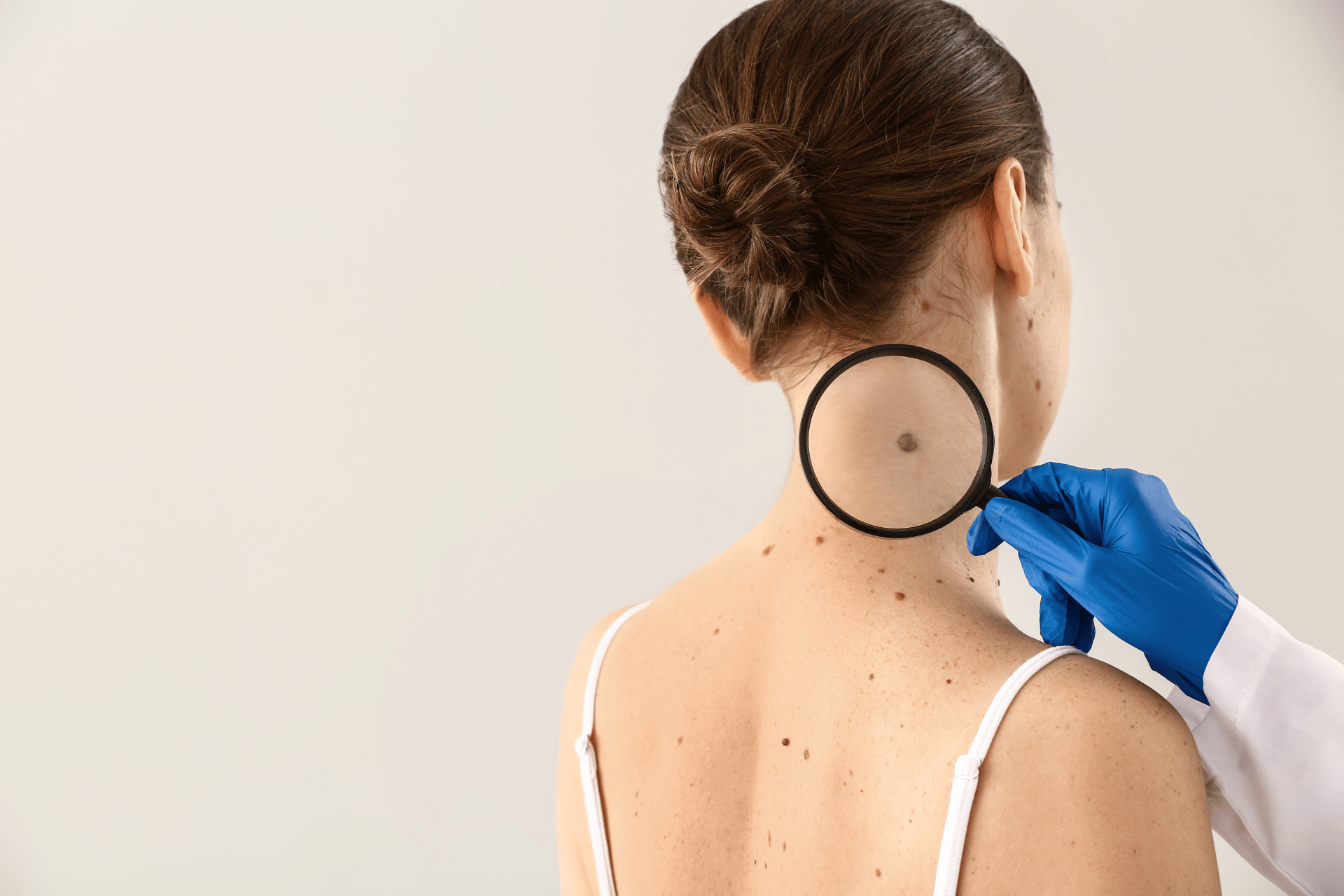
Nodular melanoma typically presents as a round, dome-shaped bump that rises clearly above the surrounding skin. Many cases are dark, appearing black, dark brown, or bluish-black. However, a significant portion are amelanotic, meaning they have little to no dark pigmentation. Amelanotic nodular melanomas may look pink, red, skin-colored, or even white, which frequently leads patients and even clinicians to mistake them for a cyst, dermatofibroma, or other benign growth.
The lesion's surface may be smooth or ulcerated. It can bleed easily when touched or scratched. Some nodules are surrounded by a reddish halo or appear inflamed. The absence of typical melanoma warning signs is precisely what makes the amelanotic form so dangerous.
The same risk factors that apply to melanoma broadly also apply to nodular melanoma:
Ultraviolet radiation exposure is the single biggest modifiable risk factor. Repeated sunburns, prolonged unprotected sun exposure, and tanning bed use all increase risk. Notably, nodular melanoma can appear on areas of the body that receive less sun exposure, including the trunk, back, and scalp, as well as chronically sun-exposed areas.
Fair skin, light eyes, and light hair reduce the skin's ability to block UV radiation. People who freckle easily rather than tan are at elevated risk.
Family history matters significantly. Having a first-degree relative with melanoma roughly doubles a person's risk.
A personal history of prior melanoma or numerous atypical moles (dysplastic nevi) increases the likelihood of developing another melanoma, potentially of the nodular type.
Age and sex are also factors. Nodular melanoma is more common in men and tends to appear later in life, though it can occur at any age.
When a suspicious lesion is identified, the diagnostic process begins with an excisional biopsy, where the entire growth is surgically removed and sent to a pathologist for examination. This is preferred over shave or punch biopsies, which may not capture the full depth of the tumor.
The most critical measurement from the biopsy report is Breslow thickness, which measures how deep the melanoma has grown into the skin in millimeters. This single number is the strongest predictor of outcome:
Less than 1mm: five-year survival rates are generally excellent
1-4mm: intermediate risk; sentinel lymph node biopsy is typically recommended
Greater than 4mm: associated with higher risk of spread and worse survival outcomes
Additional staging involves imaging (CT, PET scan, or MRI) to determine whether the cancer has spread to lymph nodes or distant organs. The pathologist will also report on ulceration, mitotic rate, and other features that refine the prognosis and guide treatment decisions.
Understanding your melanoma treatment options in detail requires working closely with an oncology team, but being informed before those conversations helps significantly.
Surgery remains the primary treatment for localized nodular melanoma. Wide surgical excision removes the tumor along with a margin of healthy tissue around it. The size of the margin depends on Breslow thickness: thinner tumors require smaller margins; thicker tumors require wider excision.
For melanomas that have spread to nearby lymph nodes or carry high-risk features, additional treatment is typically recommended. As of 2026, the standard of care for advanced or metastatic melanoma has been transformed by immunotherapy and targeted therapy.
Combination immunotherapy using nivolumab (a PD-1 inhibitor) plus relatlimab (a LAG-3 inhibitor) has demonstrated improved survival for patients with advanced melanoma compared to single-agent immunotherapy. This combination works by releasing the brakes on the immune system, allowing it to recognize and attack melanoma cells more effectively.
Targeted therapy is an option for patients whose tumors carry BRAF mutations (approximately 50% of melanomas). BRAF and MEK inhibitor combinations can produce rapid tumor shrinkage, though resistance often develops over time. These targeted therapies are frequently used alongside immunotherapy as part of a combined strategy.
Radiation therapy may be used in specific situations, such as treating brain metastases or areas where surgery is not feasible. Patients with stage III or IV disease are often enrolled in clinical trials, which continue to advance outcomes.
No strategy eliminates the risk of nodular melanoma entirely, but several measures meaningfully reduce it:
Protective clothing provides the most reliable barrier against UV radiation. Long sleeves, wide-brimmed hats, and UV-blocking fabrics are more effective than sunscreen alone for extended outdoor exposure.
Seeking shade during peak UV hours (typically 10 a.m. to 4 p.m.) significantly reduces cumulative exposure.
Broad-spectrum sunscreen with SPF 30 or higher should be applied generously to exposed skin and reapplied every two hours, or more often if swimming or sweating. Sunscreen that is not reapplied provides far less protection than the label suggests.
Tanning beds should be avoided entirely. Indoor tanning increases melanoma risk substantially, particularly for people who begin using them before age 35.
Because nodular melanoma grows quickly, the window for early intervention is narrow. Regular skin surveillance is critical, particularly for people with elevated risk.
Monthly self-exams allow you to notice new or changing spots on areas you can easily see. Use a full-length mirror, a hand mirror for hard-to-see areas, and good lighting. Note any new bump, dome-shaped growth, or area that bleeds without injury.
Annual professional skin exams by a dermatologist are essential for high-risk individuals. A dermatologist using a dermatoscope can evaluate lesions at a level of detail not visible to the naked eye. Those with a personal or family history of melanoma, many atypical moles, or a history of significant sun exposure should not skip these appointments.
For context on how melanoma types compare and what to watch for across the skin, the article on early-stage melanoma covers detection strategies in more depth.
If you notice a new firm, raised bump that is growing quickly, do not wait and watch. The speed of nodular melanoma means that time between first noticing a lesion and seeking evaluation can have a direct impact on outcomes.
Nodular melanoma typically appears as a dome-shaped, firm bump on the skin. Many cases are dark brown, black, or bluish-black. However, a significant number are amelanotic, meaning they lack dark pigmentation and may look pink, red, or skin-colored. This can make them easy to confuse with a cyst or other benign growth.
Nodular melanoma can double in size within weeks to a couple of months. This makes it one of the fastest-growing skin cancers. Because it grows vertically into the skin rather than spreading horizontally across the surface, it can reach significant depth before it becomes highly visible.
Yes. While UV exposure is a major risk factor, nodular melanoma can develop on areas that receive less sun exposure, including the trunk, back of the scalp, and covered areas of the body. Regular full-body skin exams help catch lesions in these locations.
Breslow thickness measures how deeply a melanoma has grown into the skin, in millimeters. It is the single most important prognostic indicator in melanoma. Tumors under 1mm generally carry excellent survival rates; those over 4mm are associated with a higher likelihood of spread and worse outcomes.
When detected at an early stage and before it has spread, nodular melanoma can often be treated successfully with surgery alone. Prognosis depends heavily on Breslow thickness and whether the cancer has spread to lymph nodes or other organs. Advanced cases are increasingly managed with combination immunotherapy and targeted therapy, which have significantly improved survival rates.
Anyone with a family history of melanoma, a personal history of melanoma or atypical moles, fair skin, a history of significant UV exposure, or previous sunburns should see a dermatologist annually. People without these risk factors benefit from annual self-exams and periodic professional exams starting in their 30s.
Nodular melanoma is rare but moves faster than almost any other skin cancer. Its tendency to grow downward rather than spread outward, its ability to appear without obvious pigmentation, and its rapid pace of growth make it easy to overlook until it has already reached a dangerous stage. Understanding the EFG rule, knowing your personal risk factors, and committing to regular self-exams and professional screenings are the most practical tools available for catching it early.
If you notice a new, raised, firm bump that is growing quickly, contact a dermatologist promptly. Doctronic.ai can help you evaluate symptoms and connect with a specialist when time matters.
Understanding Accutane and Breastfeeding SafetyAccutane, also known by its generic name isotretinoin, is a powerful oral medication used to treat severe acne that hasn't [...]
Read MoreUnderstanding Imiquimod and Pregnancy Safety ClassificationsImiquimod, commonly known by the brand name Aldara, carries an FDA pregnancy category C classification, which [...]
Read MoreUnderstanding Imiquimod Drug InteractionsImiquimod cream works by stimulating your body's immune system to fight abnormal skin cells, making it essential to understand [...]
Read More
Join 50,000+ readers using Doctronic to understand symptoms, medications,
and next steps.
Add your phone number below to get health updates and exclusive VIP offers.
By providing your phone number, you agree to receive SMS updates from Company. Message and data rates may apply. Reply “STOP” to opt-out anytime. Read our Privacy Policy and Terms of Service for more details.
Save your consults. Talk with licensed doctors and manage your health history.