
Differin (Adapalene) for Seniors: What to Know
Read More

Medically reviewed by Faith Coleman , University of New Mexico School of Medicine on May 26th, 2026. Updated on May 28th, 2026
Most brown spots on the skin are harmless and fall into a small number of well-defined categories: solar lentigines (sunspots), seborrheic keratoses, melasma, or post-inflammatory hyperpigmentation.
Solar lentigines are flat, clearly defined brown spots on sun-exposed areas that result from years of UV exposure.
Seborrheic keratoses are rough, wart-like brown growths that are benign but can be mistaken for something more serious.
The ABCDE rule (Asymmetry, Border, Color, Diameter, Evolving) is the standard first check for distinguishing a harmless brown spot from a potentially cancerous one.
Any brown spot that is new, changing, bleeding, or growing rapidly should be evaluated by a dermatologist regardless of how it looks.
For a quick assessment of a concerning skin spot, Doctronic.ai connects you with licensed physicians through free AI consultations and affordable telehealth visits available any time.
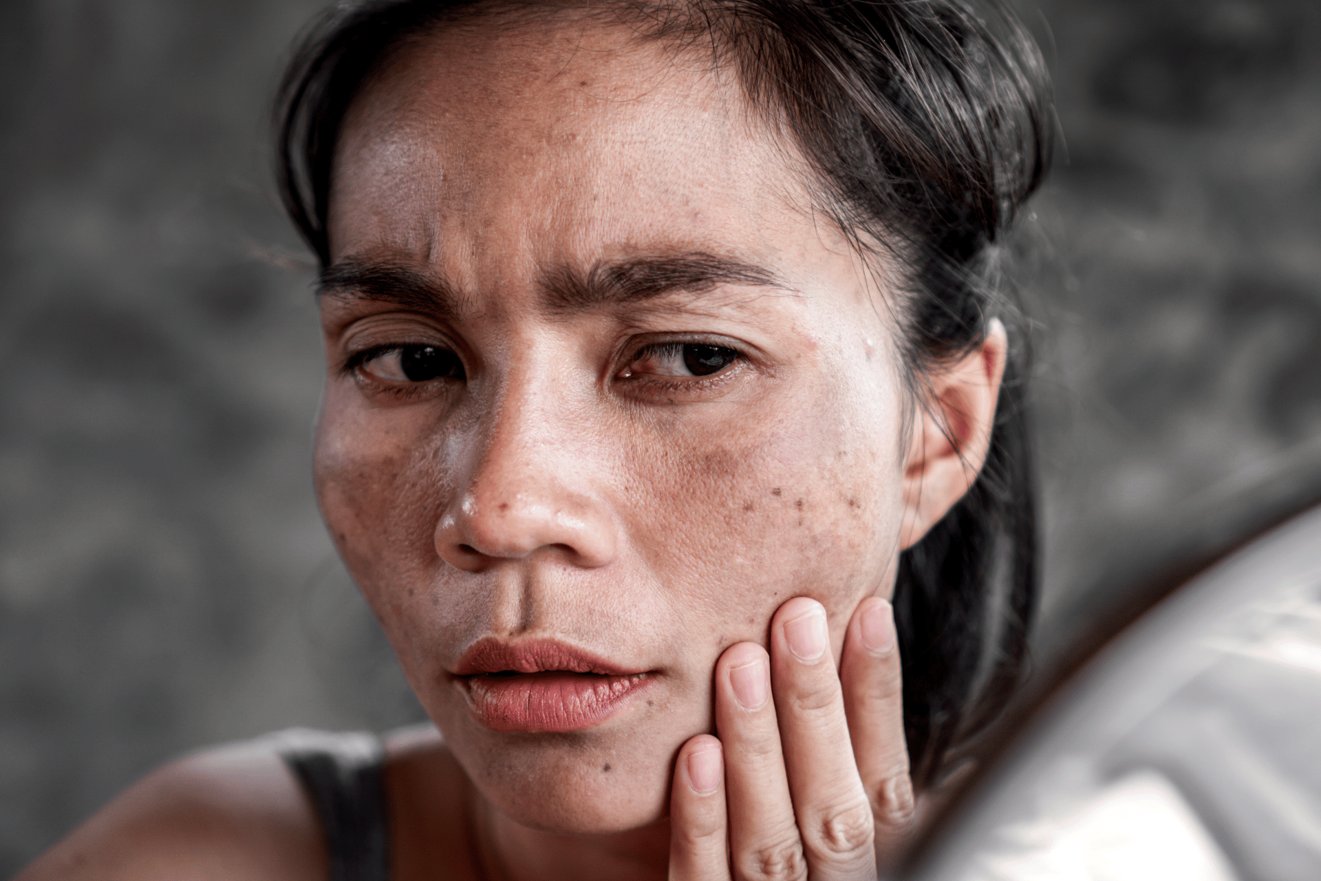
Skin accumulates the effects of decades of UV exposure, hormonal changes, inflammation, and routine cell turnover, and brown spots are among the most visible results of that history. The vast majority are harmless. A small subset requires attention.
The most useful approach to a new brown spot is not to try to diagnose it from memory or by matching it to a photo, but to understand which features separate the harmless from the concerning, and when a professional evaluation is appropriate.
Solar lentigines, commonly called sunspots, age spots, or liver spots, are the most frequently encountered brown spots on sun-exposed skin. They appear in areas where cumulative UV exposure has been highest: face, hands, forearms, shoulders, and decolletage.
Sunspots are flat, have cistinct boundaries and a uniform brown color. They range from a few millimeters to about a centimeter. They do not have the raised, rough texture of seborrheic keratoses. They develop gradually over years and do not change rapidly.
Solar lentigines result from melanocytes overproducing melanin locally in response to chronic UV exposure. They are not precancerous, but their presence indicates that cumulative sun damage has occurred, which is associated with an elevated overall skin cancer risk. Age spots vs. sun spots are sometimes distinguished by cause (hormonal vs. UV), but the terms are often used interchangeably in everyday language.
Seborrheic keratoses are benign skin growths that appear, usually starting in middle age, as brown, black, or tan patches with a rough, waxy, or "stuck-on" texture. They can look unsettling because they grow over time, can become darker, and sometimes resemble melanoma to untrained eyes.
Seborrheic keratoses are benign and have no relationship to skin cancer. However, a seborrheic keratosis that has recently changed significantly, bleeds without trauma, or looks atypical should be evaluated by a dermatologist to confirm the diagnosis.
Common features: rough or warty surface texture, slightly elevated above the skin surface, well-defined borders, and a tan to dark brown color that appears almost applied to the surface rather than originating from within.
Melasma produces larger, irregular brown or gray-brown patches, most commonly on the cheeks, forehead, upper lip, and chin. It is driven by a combination of UV exposure and hormonal influences, which is why it frequently appears during pregnancy, while using hormonal contraceptives, or at perimenopause.
Melasma is symmetrical (both sides of the face are usually affected similarly), which helps distinguish it from solar lentigines and from asymmetric lesions that might warrant concern. It is benign but can be persistent and difficult to treat without consistent sun protection.
Post-inflammatory hyperpigmentation (PIH) is the brown mark left behind after the skin heals from an inflammatory event: acne, an insect bite, a minor wound, eczema, or any other condition that caused inflammation. The melanocytes in affected areas overproduce pigment during the healing process, leaving a flat brown spot that can persist for months.
PIH is most common in people with medium to deep skin tones, where melanocytes are more reactive to inflammation. Unlike solar lentigines, PIH appears wherever the triggering event occurred rather than specifically on sun-exposed areas.
Skin cancer pictures can help illustrate what concerning lesions look like, but visual comparison is less reliable than the ABCDE rule for initial self-assessment.
Asymmetry: one half of the spot does not match the other.
Border: the edges are irregular, jagged, notched, or blurred.
Color: the spot contains multiple shades of brown, red, white, or black rather than a single uniform color.
Diameter: larger than 6 millimeters (about the size of a pencil eraser) warrants attention, though smaller melanomas exist.
Evolving: any change in size, color, shape, or new symptoms like bleeding, itching, or crusting.
Any brown spot that meets one or more of these criteria, or that is new and unexplained on a person with significant sun exposure history, should be evaluated by a dermatologist.
Annual skin checks with a dermatologist are also appropriate for anyone with multiple moles, a personal or family history of skin cancer, or significant cumulative sun exposure.
Brown spots are not automatically signs of cancer, liver disease (despite the colloquial name "liver spots," they have no connection to liver function), or aging disease. They are not contagious. Solar lentigines and seborrheic keratoses, which together account for the vast majority of brown spots adults notice, are benign findings that need no treatment unless desired for cosmetic reasons.
The exception to this pattern is any brown spot that changes quickly, has irregular features, or appears in an unusual location like a nail bed or palm.
New spots rarely appear truly overnight; what usually happens is that a spot that was gradually developing becomes noticeable suddenly. True rapid-onset brown marks after trauma (bruising) or inflammation (PIH) can appear within days. A genuinely new spot that appears within days and has no clear cause should be evaluated.
Most brown spots (solar lentigines, seborrheic keratoses, melasma, PIH) do not become cancerous. Solar lentigines indicate UV damage but are not precancerous lesions. However, certain lesions called dysplastic nevi (atypical moles) can progress to melanoma, which is why professional evaluation of any changing or atypical lesion matters.
Over-the-counter brightening ingredients like niacinamide, vitamin C, and kojic acid can gradually fade solar lentigines and PIH over weeks to months. Prescription-strength options and professional treatments (laser, chemical peel, cryotherapy) produce faster results. Seborrheic keratoses do not respond to topical brighteners. No home treatment should be used on a spot that has not been evaluated and confirmed benign.
Cumulative UV exposure is the primary driver for solar lentigines and some seborrheic keratoses. Over decades, specific melanocytes accumulate enough UV-induced signaling to begin producing excess pigment locally. This is why brown spots appear in greatest concentration on the most sun-exposed areas and accumulate with age.
Moles (nevi) are formed by clusters of melanocytes and are typically raised, uniformly colored, and present from early in life. Solar lentigines and seborrheic keratoses are different in origin and structure. The distinction matters clinically because moles carry different cancer risk profiles than these other benign lesions.
Most brown spots on the skin are harmless. Solar lentigines from UV exposure, seborrheic keratoses, melasma, and post-inflammatory marks account for the overwhelming majority. The ABCDE framework is the most practical tool for initial self-assessment, and any spot that is asymmetric, has irregular borders, contains multiple colors, is growing, or is changing should be evaluated by a dermatologist. When in doubt, a professional skin check provides certainty that visual self-assessment cannot. For fast access to a licensed physician who can assess a skin concern, Doctronic.ai offers affordable telehealth visits available any time.

Join 50,000+ readers using Doctronic to understand symptoms, medications,
and next steps.
Add your phone number below to get health updates and exclusive VIP offers.
By providing your phone number, you agree to receive SMS updates from Company. Message and data rates may apply. Reply “STOP” to opt-out anytime. Read our Privacy Policy and Terms of Service for more details.
Save your consults. Talk with licensed doctors and manage your health history.