
Clarinex (Desloratadine) Storage and Expiration: Does It Still Work?
Understanding Desloratadine Expiration DatesDesloratadine, the active ingredient in Clarinex, follows FDA requirements for expiration date testing and labeling. [...]
Read More
Medically reviewed by Veronica Hackethal | MD, MSc , Harvard University | University of Oxford | Columbia Vagelos College of Physicians and Surgeons on May 25th, 2026. Updated on May 26th, 2026
A typical mole is not cancerous, but having many moles slightly increases melanoma risk rather than each typical mole being a direct signal of higher risk
People with more than five dysplastic nevi face roughly 10 times the melanoma risk compared to those without any
About 1 in 10 Americans have at least one atypical mole, making regular monitoring essential
The ABCDE method helps identify suspicious moles: Asymmetry, Border irregularity, Color variation, Diameter, and Evolution
Monthly self-examinations combined with annual professional skin checks provide the best protection
Doctronic.ai offers accessible AI-powered consultations to help track skin changes and determine when professional evaluation is needed
Finding an unusual-looking mole can trigger immediate worry. The question of whether atypical moles are cancerous deserves a clear answer: no, they are not cancer. These moles are benign growths that look different from typical moles. The concern arises because having atypical moles indicates a higher chance of developing melanoma later. Understanding this risk helps people take appropriate action without unnecessary panic. Doctronic.ai can help evaluate skin concerns and guide decisions about when to seek in-person care.
A dysplastic nevus is the medical term for an atypical mole. These moles share some visual characteristics with melanoma but remain benign. They typically appear larger than regular moles, often exceeding 5 millimeters in diameter. Their color tends to be uneven, mixing shades of tan, brown, and pink within a single mole. The edges often look irregular or fade into surrounding skin rather than having crisp borders. About 1 in 10 Americans have at least one atypical mole, making this a common finding during skin examinations.
Benign moles pose no health threat and require no treatment beyond monitoring. Precancerous lesions show cellular changes that could progress to cancer without intervention. Malignant growths are active cancers requiring immediate treatment. Atypical moles fall into a gray zone: they are benign but serve as markers for elevated melanoma risk. The mole itself rarely transforms into cancer. The real concern is that having these moles indicates skin that may be prone to developing melanoma elsewhere.
Dermatologists developed the ABCDE method to help identify concerning moles. Asymmetry means one half does not match the other. Border irregularity shows as ragged, notched, or blurred edges. Color variation includes multiple shades within a single mole. Diameter larger than a pencil eraser raises concern. Evolution refers to any change in size, shape, color, or symptoms like itching or bleeding. Any mole meeting multiple criteria deserves professional evaluation.
Atypical moles appear most frequently on areas that receive intermittent sun exposure rather than only chronically sun-exposed sites. The back, chest, and legs are common sites. They can also develop in areas with intermittent sun exposure, such as the scalp and buttocks. Women often find them on their legs, while men frequently discover them on their backs. Checking hard-to-see areas requires a mirror or assistance from a partner.
Family history significantly influences melanoma risk. Having a first-degree relative with melanoma doubles or triples personal risk. Some families carry mutations in genes like CDKN2A that dramatically increase susceptibility. Familial atypical multiple mole melanoma syndrome affects families with numerous dysplastic nevi and melanoma history. Genetic counseling may benefit those with strong family histories.
Ultraviolet radiation damages skin cell DNA over time. Blistering sunburns during childhood and adolescence particularly increase melanoma risk. Use of indoor tanning devices before age 35 has been associated with a 59 percent increase in melanoma risk, based on the latest meta-analyses. Cumulative sun exposure matters too, not just severe burns. Fair-skinned individuals burn more easily and face higher risk, though melanoma affects all skin types.
The number of moles on the body directly correlates with melanoma risk. People with more than 100 common moles face elevated risk. Those with more than five dysplastic nevi face a melanoma risk about 10 times greater than someone without any. Counting moles provides valuable information for determining monitoring frequency and protective measures.
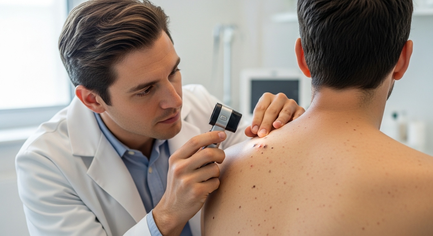
Annual full-body skin examinations by a dermatologist remain the gold standard for monitoring. Early mole screening for dysplastic nevi is crucial, and experts recommend total cutaneous examination, total cutaneous photography, and dermoscopy for managing these lesions. Dermoscopy uses a specialized magnifying device to examine moles in detail. This technique reveals patterns invisible to the naked eye and helps distinguish benign moles from suspicious ones.
Dermatologists recommend biopsy when a mole shows concerning features or changes over time. The procedure involves removing all or part of the mole for microscopic examination. Pathologists grade dysplastic nevi as mild, moderate, or severe based on cellular abnormalities. Mildly dysplastic moles rarely require further treatment if completely removed. Severely dysplastic moles may need wider excision to ensure complete removal.
High-risk individuals benefit from total body photography. This technique creates a baseline record of all moles. During follow-up visits, dermatologists compare current appearance to photographs. New moles or changes become immediately apparent. Some clinics offer digital dermoscopy with computer-assisted analysis to track subtle changes over time. Doctronic.ai can help individuals understand their risk profile and determine appropriate monitoring schedules.
Monthly self-examinations catch changes between professional visits. Choose a consistent day each month to check the entire body. Use a full-length mirror and hand mirror to see all areas. Photograph moles with a smartphone to track changes over time. Pay attention to any mole that looks different from others or changes in any way.
A systematic approach ensures no areas are missed:
Start with the face, ears, and scalp
Check arms, including underarms and palms
Examine the torso front and back
Inspect legs, including between toes and soles
Use mirrors for the back and buttocks
Consistent sun protection reduces the development of new moles and lowers melanoma risk. Broad-spectrum sunscreen with SPF 30 or higher should be applied daily, not just at the beach. Reapplication every two hours during outdoor activities maintains protection. Protective clothing, including wide-brimmed hats and UV-blocking sunglasses, provides physical barriers.
Seeking shade during peak UV hours between 10 AM and 4 PM limits exposure. Window film on car windows blocks UV rays during commutes. These habits become especially important for those with multiple atypical moles or family melanoma history.
Atypical moles rarely transform directly into melanoma. The greater concern is that having these moles indicates skin prone to developing melanoma independently. Regular monitoring catches any problems early when treatment is most effective.
Most dermatologists recommend annual full-body examinations for people with atypical moles. Those with numerous dysplastic nevi or family melanoma history may need examinations every three to six months.
Removing every atypical mole is neither necessary nor practical for most people. Dermatologists typically recommend removal only for moles showing concerning changes or severe dysplasia on biopsy.
Having atypical moles does not guarantee melanoma development. Many people with dysplastic nevi never develop skin cancer. Proper monitoring and sun protection significantly reduce risk.
Atypical moles typically appear during puberty and continue developing into the 30s and 40s. Children can develop them, particularly those with fair skin or family history of dysplastic nevi.
Atypical moles are not cancerous but signal increased melanoma risk requiring vigilant monitoring. Monthly self-examinations combined with annual professional skin checks provide the best protection for long-term skin health. For personalized guidance on tracking skin changes and understanding when to seek professional evaluation, visit Doctronic.ai for accessible AI-powered medical consultations available around the clock.
Understanding Desloratadine Expiration DatesDesloratadine, the active ingredient in Clarinex, follows FDA requirements for expiration date testing and labeling. [...]
Read MoreThe Science Behind Diazepam and Hair LossDiazepam (Valium) belongs to the benzodiazepine class of medications, primarily prescribed for anxiety, muscle spasms, and seizure [...]
Read MoreUnderstanding Quviviq Storage RequirementsQuviviq (daridorexant) requires specific storage conditions to maintain its therapeutic effectiveness for treating insomnia. This [...]
Read More
Join 50,000+ readers using Doctronic to understand symptoms, medications,
and next steps.
Add your phone number below to get health updates and exclusive VIP offers.
By providing your phone number, you agree to receive SMS updates from Company. Message and data rates may apply. Reply “STOP” to opt-out anytime. Read our Privacy Policy and Terms of Service for more details.
Save your consults. Talk with licensed doctors and manage your health history.