
Clarinex (Desloratadine) Storage and Expiration: Does It Still Work?
Understanding Desloratadine Expiration DatesDesloratadine, the active ingredient in Clarinex, follows FDA requirements for expiration date testing and labeling. [...]
Read More
Medically reviewed by Veronica Hackethal | MD, MSc , Harvard University | University of Oxford | Columbia Vagelos College of Physicians and Surgeons on May 25th, 2026. Updated on May 26th, 2026
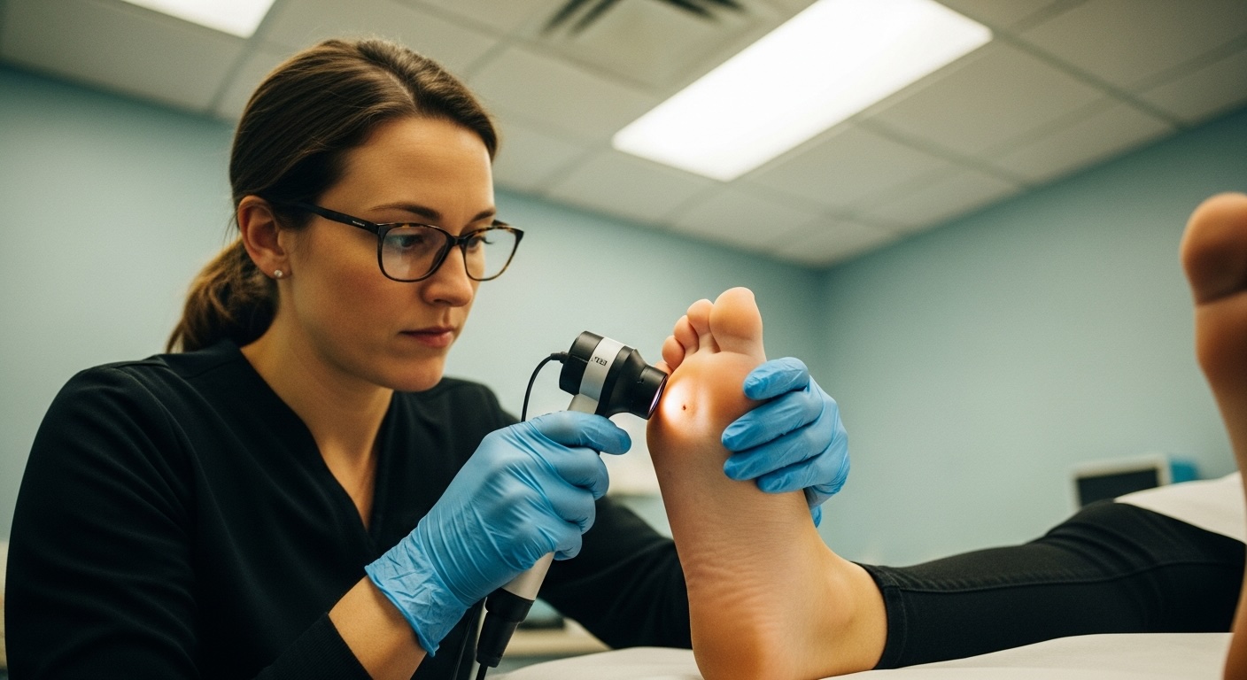
Acral lentiginous melanoma (ALM) develops on palms, soles, and under nails, making it the skin cancer most often missed during routine screenings
ALM accounts for only 1-3% of all melanoma cases but represents up to 70% of melanomas in people with darker skin tones
Misdiagnosis rates range between 20% and 34% because ALM mimics bruises, warts, and fungal infections
The CUBED acronym helps with early detection: Color irregularity, Uncertain diagnosis, Bleeding, Enlarged over 7mm, and Delay in healing
Regular self-examinations of hands, feet, and nails can catch ALM before it spreads
If you notice suspicious changes on your palms, soles, or nails, Doctronic.ai offers 24/7 telehealth consultations to help you determine your next steps
A dark streak appears under a toenail. Most people assume they stubbed it or dropped something heavy on their foot. Months pass before anyone thinks to mention it to a doctor. This delay can prove fatal. Acral lentiginous melanoma is the skin cancer most often missed because it hides in plain sight on body parts people rarely examine: the palms of hands, soles of feet, and beneath fingernails and toenails. Unlike other melanomas linked to sun exposure, ALM strikes regardless of UV history. This makes traditional skin cancer awareness campaigns nearly useless for catching it early. Understanding where to look and what to look for saves lives.
ALM develops in the acral regions of the body, the hairless skin of palms, soles, and the nail matrix. As a rare skin cancer, it requires specific awareness. The cancer begins in melanocytes, the cells responsible for skin pigment. Unlike superficial spreading melanoma, ALM grows in a lentiginous pattern, meaning it spreads horizontally through the epidermis before invading deeper tissues. This horizontal growth phase can last months or years, providing a window for early detection that too many people miss.
ALM accounts for only 1-3% of all melanoma cases diagnosed in Caucasian populations. Among Black, Asian, and Hispanic individuals, this percentage jumps dramatically, representing 30-70% of all acral melanoma diagnoses in these groups. The reason is straightforward: people with more melanin have natural protection against UV-induced melanomas but remain equally vulnerable to ALM, which develops independently of sun exposure.
Traditional melanoma prevention focuses on sunscreen, avoiding tanning beds, and limiting UV exposure. None of these measures prevent ALM. The genetic mutations driving ALM differ from those in sun-related melanomas. While UV protection remains important for other skin cancers, it does not specifically reduce ALM risk. This disconnect between public health messaging and ALM reality contributes to delayed diagnoses. Patients and doctors alike fail to consider skin cancer on body parts that rarely see sunlight.
A longitudinal pigmented band running from the nail base to the tip demands immediate attention. While benign causes exist, especially in people with darker skin who commonly have melanonychia striata, certain characteristics signal danger. Bands wider than 3mm, bands that suddenly appear in adulthood, and bands affecting a single digit rather than multiple nails require evaluation. Doctronic.ai can help users understand these warning signs and determine when professional evaluation is necessary.
Medical experts developed a practical screening tool specifically for acral lesions. The CUBED acronym identifies high-risk features: Color irregularity with multiple shades, Uncertain diagnosis by initial examination, Bleeding from the lesion, Enlarged diameter over 7mm, and Delay in healing beyond two months. Any lesion meeting two or more criteria warrants biopsy.
When pigment from a nail streak extends onto the surrounding skin fold, called the proximal nail fold, doctors recognize this as Hutchinson's sign. This finding strongly suggests subungual melanoma rather than benign causes. The pigment spreads because cancer cells migrate beyond the nail matrix into adjacent tissue. Pseudo-Hutchinson's sign can occur with benign conditions, but true Hutchinson's sign requires urgent biopsy.
ALM faces significant diagnostic challenges, with misdiagnosis rates ranging between 20% and 34%, far exceeding rates for other melanoma subtypes. Plantar ALM resembles warts, calluses, or diabetic ulcers. Subungual melanoma gets treated as fungal infection for months before anyone considers biopsy. Amelanotic variants lacking pigment look like non-healing wounds. Each misdiagnosis delays treatment and allows deeper invasion.
Medical training historically emphasized melanoma detection on fair skin. Dermatology textbooks underrepresented images of skin conditions in people of color. This educational gap means clinicians may feel less confident examining darker skin or may not immediately consider melanoma in these patients. The result: later-stage diagnoses and worse outcomes for the populations most affected by ALM.
Dermoscopy uses magnification and polarized light to examine skin lesions in detail. On acral skin, benign nevi display a parallel furrow pattern, with pigment following the skin creases. ALM shows a parallel ridge pattern, with pigment concentrated on the ridges between furrows. This distinction helps dermatologists identify suspicious lesions before biopsy. Doctronic.ai provides educational resources to help patients understand what dermoscopy findings mean.
Visual examination alone cannot diagnose melanoma. Suspicious acral lesions require tissue sampling. Punch biopsies extract a small cylinder of tissue for histological analysis. For nail lesions, nail matrix biopsy may be necessary. The pathology report determines melanoma thickness, ulceration status, and mitotic rate, all factors that guide staging and treatment planning.
Early-stage ALM requires wide local excision with margins determined by tumor thickness. On acral surfaces, achieving adequate margins while preserving function presents challenges. Mohs micrographic surgery, which examines tissue margins during the procedure, maximizes tissue preservation while ensuring complete tumor removal. Mohs surgery is not yet a standard treatment for all ALM cases but may be selectively used for in situ or very early lesions where tissue preservation is critical. Some cases require digit amputation when the tumor involves bone or when margins cannot otherwise be achieved.
When ALM spreads beyond the primary site, immunotherapy offers hope. Checkpoint inhibitors like pembrolizumab and nivolumab help the immune system recognize and attack melanoma cells. Targeted therapies work for tumors with specific mutations. Response rates for metastatic ALM have improved significantly with these treatments, though outcomes still lag behind other melanoma subtypes. Combination regimens and novel agents, such as relatlimab with nivolumab, have shown enhanced efficacy for advanced disease. Regular monitoring through platforms like Doctronic.ai helps patients track symptoms and communicate effectively with their oncology teams.
Monthly self-examinations should include areas traditional skin checks ignore. Examine the soles of both feet using a mirror or ask a partner for help. Check between toes. Look at palms and the spaces between fingers. Inspect each fingernail and toenail, noting any new pigmented bands or changes to existing ones. Document suspicious findings with photographs to track changes over time.
People with risk factors need heightened vigilance. Family history of melanoma, personal history of any skin cancer, and immunosuppression all increase risk. Those with many benign moles on acral surfaces should establish baseline documentation with a dermatologist.
ALM rarely affects children, but it can occur. Any pigmented band on a child's nail or any unusual spot on palms or soles deserves evaluation, especially if it changes over time.
The horizontal growth phase may last one to several years before vertical invasion begins. Once ALM invades deeper tissues, metastasis can occur within months. Early detection during the horizontal phase dramatically improves survival.
Genetic predisposition plays a role in some cases. Families with multiple melanoma diagnoses or those carrying certain gene mutations face elevated risk. Genetic counseling may benefit high-risk families.
People who work barefoot or experience repeated trauma to hands and feet may face slightly elevated risk, though research remains inconclusive. The trauma theory suggests chronic injury may trigger melanocyte changes.
Acral lentiginous melanoma hides where people least expect skin cancer: on palms, soles, and under nails. Regular self-examination of these areas catches ALM early when treatment succeeds. For questions about suspicious spots or concerning symptoms, Doctronic.ai offers free AI doctor visits and affordable telehealth consultations available 24/7.
Understanding Desloratadine Expiration DatesDesloratadine, the active ingredient in Clarinex, follows FDA requirements for expiration date testing and labeling. [...]
Read MoreThe Science Behind Diazepam and Hair LossDiazepam (Valium) belongs to the benzodiazepine class of medications, primarily prescribed for anxiety, muscle spasms, and seizure [...]
Read MoreUnderstanding Quviviq Storage RequirementsQuviviq (daridorexant) requires specific storage conditions to maintain its therapeutic effectiveness for treating insomnia. This [...]
Read More
Join 50,000+ readers using Doctronic to understand symptoms, medications,
and next steps.
Add your phone number below to get health updates and exclusive VIP offers.
By providing your phone number, you agree to receive SMS updates from Company. Message and data rates may apply. Reply “STOP” to opt-out anytime. Read our Privacy Policy and Terms of Service for more details.
Save your consults. Talk with licensed doctors and manage your health history.