
Dysplastic Nevi and Skin Cancer Risk: What You Need to Know
What You Should Know About Atypical MolesFinding an unusual mole can be alarming. The good news: most dysplastic nevi never become cancerous. The challenging news: having [...]
Read More
Medically reviewed by Alan Lucks | MD , Alan Lucks MDPC Private Practice - New York on May 22nd, 2026. Updated on May 22nd, 2026
Dysplastic nevi are atypical moles with irregular features that differ from common moles in size, color, and border characteristics.
People with five or more atypical moles are estimated to be about six times more likely to develop melanoma compared to those without them.
Most melanomas in people with dysplastic nevi actually arise on normal-appearing skin, not from the atypical moles themselves.
Regular dermoscopy screenings and photographic mapping help track changes in moles over time.
Self-examinations combined with professional dermatological checkups create the strongest defense against skin cancer progression.
Doctronic.ai offers AI-powered consultations to help individuals understand mole changes and determine when to schedule a dermatology appointment.
Finding an unusual mole can be alarming. The good news: most dysplastic nevi never become cancerous. The challenging news: having them signals an elevated risk for melanoma that requires attention.
Understanding dysplastic nevi and skin cancer risk helps people make informed decisions about monitoring and prevention. The lifetime risk of developing melanoma is approximately 2.6% for non-Hispanic White individuals in the United States, but this percentage climbs significantly for those with multiple atypical moles or a family history of melanoma.
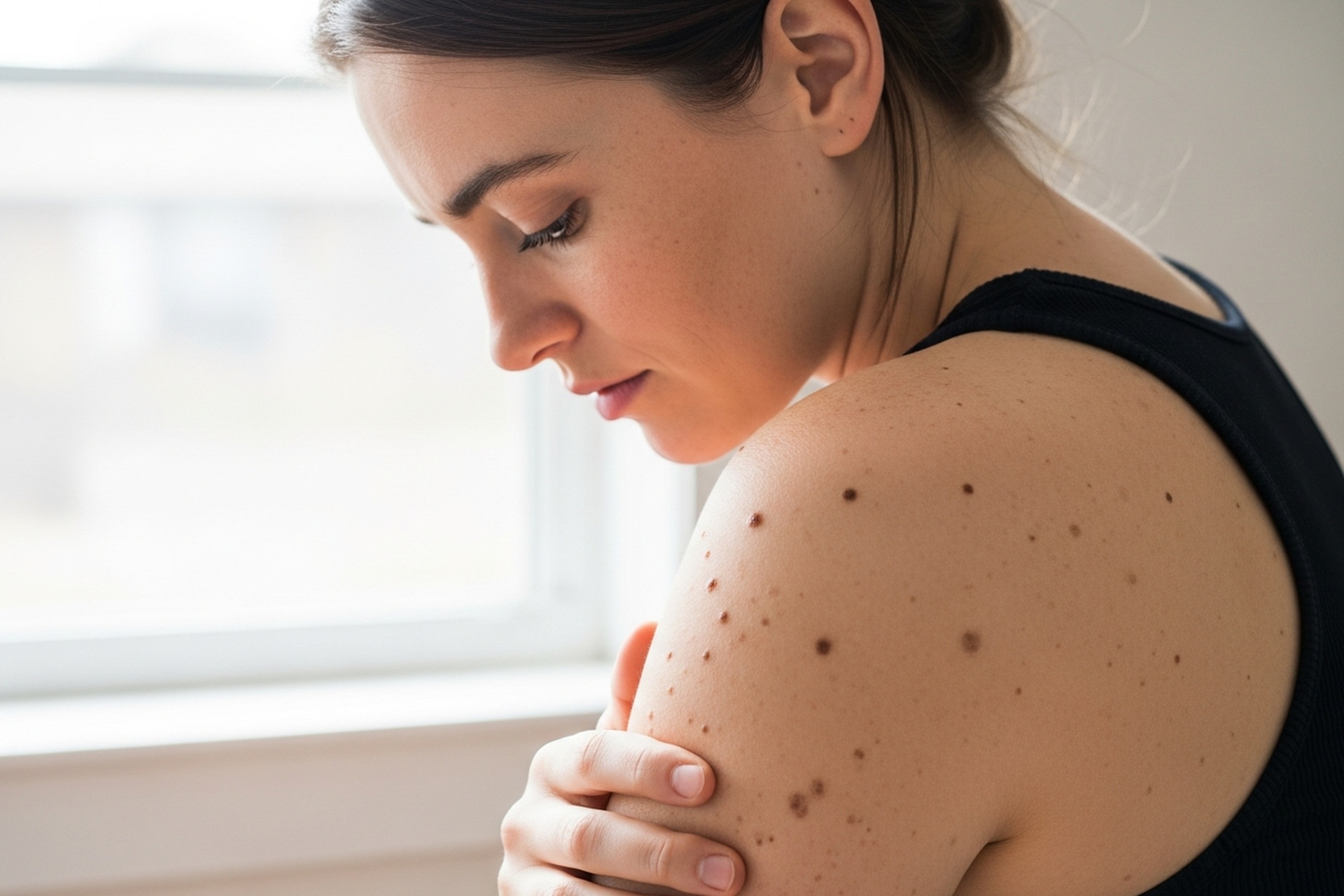
Dysplastic nevi look different from ordinary moles. They tend to be larger, often exceeding 5 millimeters in diameter. Their borders appear fuzzy or irregular rather than smooth and well-defined. Color varies within a single mole, mixing shades of tan, brown, pink, and sometimes red.
Dermatologists use the ABCDE criteria to evaluate moles:
Asymmetry: One half doesn't match the other.
Border irregularity: Notched, scalloped, or blurred edges.
Color variation: Multiple shades within the same mole.
Diameter: Greater than 6 millimeters raises concern.
Evolution: Any change in size, shape, color, or symptoms like itching or bleeding.
Common moles are typically uniform in color, round or oval in shape, and smaller than a pencil eraser. They have clear borders and stay stable over time.
Dysplastic nevi break these rules. They may appear flat with a raised center, show multiple colors, and have indistinct edges that fade into surrounding skin. A person might have one dysplastic nevus or dozens scattered across their body.
The relationship between dysplastic nevi and melanoma is complex. Having atypical moles serves as a marker for increased melanoma risk, but the moles themselves rarely transform into cancer.
Research shows that melanomas in patients with dysplastic nevi more often arise on skin without a preexisting mole, although some melanomas can develop within dysplastic nevi. This means surveillance must cover the entire skin surface, not just existing atypical moles.
Dysplastic nevus syndrome runs in families. When multiple family members have numerous atypical moles and a history of melanoma, the condition follows an inherited pattern. Genetic mutations affecting melanocyte development and DNA repair mechanisms contribute to this familial risk.
First-degree relatives of individuals with dysplastic nevus syndrome or melanoma should undergo regular dermatologic skin examinations regardless of their own mole count.
Numbers tell an important story. Having one or two dysplastic nevi slightly elevates melanoma risk. Having five or more increases it about sixfold.
The presence of atypical moles combined with a personal or family history of melanoma creates the highest risk category. These individuals benefit from the most aggressive monitoring protocols. People noticing changes in existing moles or new spots alongside a family history of melanoma should seek professional evaluation. Those unsure whether a spot is benign can learn to compare lesions through resources on identifying suspicious skin changes.
Proper diagnosis requires professional assessment. Self-examination catches changes, but clinical evaluation determines the nature of suspicious moles.
Dermoscopy uses a handheld device with magnification and polarized light to see structures beneath the skin surface. This technique reveals pigment patterns, vascular structures, and other features invisible to the naked eye.
Dermoscopy increases diagnostic accuracy and helps dermatologists distinguish benign atypical moles from early melanomas. Regular examinations create a baseline for tracking changes. Doctronic.ai can help patients prepare questions for their dermatology appointments and understand what to expect during screenings.
Biopsy becomes necessary when a mole shows concerning features or changes over time:
Excisional biopsy removes the entire mole with a margin of normal skin.
Shave biopsy removes the raised portion.
Punch biopsy takes a deeper, circular sample.
The choice depends on the mole's characteristics and location.
Pathologists grade dysplastic nevi based on how abnormal the cells appear:
Mild atypia: Slight irregularities, typically requires monitoring only.
Moderate atypia: More pronounced changes, may require re-excision if margins are positive.
Severe atypia: Approaches melanoma in situ, generally warrants wider excision.
Treatment decisions balance cancer prevention against the impracticality of removing every atypical mole. Most dysplastic nevi require monitoring rather than removal.
Moles with mild atypia and clear margins on biopsy typically need observation only. Moles that change rapidly, develop new symptoms, or cause concern for the patient deserve prompt evaluation and possible excision. Comparing moles against photos taken over time supports better self-monitoring between professional visits. Patients interested in remote evaluation options can explore telehealth dermatology care for initial assessments.
Total body photography creates a visual record of all moles. Sequential photographs taken months or years apart reveal subtle changes that might otherwise go unnoticed.
Digital dermoscopy adds magnified images of individual moles to this record. This documentation proves invaluable for patients with numerous atypical moles, allowing precise tracking of each lesion over time.
UV radiation damages DNA in skin cells and promotes melanoma development. For people with dysplastic nevi, sun protection carries special importance:
Broad-spectrum sunscreen with SPF 30 or higher on all exposed skin daily.
Protective clothing, wide-brimmed hats, and UV-blocking sunglasses.
Seeking shade during peak sun hours between 10 AM and 4 PM.
Avoiding tanning beds entirely.
Monthly self-examinations help catch changes between professional visits. A full-length mirror and hand mirror allow visualization of the entire body. Partners can help examine hard-to-see areas like the back and scalp.
Any new mole, changing mole, or mole that looks different from others warrants professional evaluation. Taking smartphone photos of concerning moles helps track changes between appointments.
People with dysplastic nevi should see a dermatologist at least annually. Those with dysplastic nevus syndrome, personal melanoma history, or family melanoma history may need examinations every three to six months.
Doctronic.ai provides 24/7 access to medical guidance that can help patients determine when symptoms warrant an urgent appointment versus routine monitoring.
Most dysplastic nevi remain stable and never become cancerous. Melanomas in people with atypical moles more commonly develop on normal-appearing skin rather than from existing dysplastic nevi. The moles serve as risk markers rather than direct cancer precursors.
No. Removing every atypical mole is impractical and unnecessary for most patients. Dermatologists recommend removal for moles showing severe atypia, rapid changes, or concerning features on dermoscopy. Stable moles with mild atypia typically require monitoring only.
Annual examinations represent the minimum for people with atypical moles. Those with dysplastic nevus syndrome, multiple melanoma risk factors, or personal melanoma history benefit from examinations every three to six months.
Having dysplastic nevi does not guarantee melanoma development. It indicates elevated risk that requires appropriate monitoring and sun protection. Many people with numerous atypical moles never develop skin cancer when they follow proper surveillance protocols.
Understanding dysplastic nevi and their connection to melanoma risk empowers people to take appropriate precautions without unnecessary anxiety. Regular professional examinations, consistent self-checks, and diligent sun protection create a strong defense against skin cancer.
For questions about atypical moles or any skin concerns, visit Doctronic.ai for AI-powered consultations that provide personalized guidance based on the latest medical research.
What You Should Know About Atypical MolesFinding an unusual mole can be alarming. The good news: most dysplastic nevi never become cancerous. The challenging news: having [...]
Read More
Join 50,000+ readers using Doctronic to understand symptoms, medications,
and next steps.
Add your phone number below to get health updates and exclusive VIP offers.
By providing your phone number, you agree to receive SMS updates from Company. Message and data rates may apply. Reply “STOP” to opt-out anytime. Read our Privacy Policy and Terms of Service for more details.
Save your consults. Talk with licensed doctors and manage your health history.